Trigger Point Therapy for Myofascial Pain (28 page)
Read Trigger Point Therapy for Myofascial Pain Online
Authors: L.M.T. L.Ac. Donna Finando

Walking is one of the best exercises for this muscle.
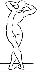
Stretch exercise 1: Gluteus minimus
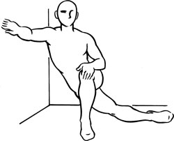
Stretch exercise 2: Gluteus minimus

Tensor fasciae latae and trigger point
T
ENSOR
F
ASCIAE
L
ATAE
Proximal attachment:
Anterior iliac crest, just posterior to the anterior superior iliac spine (ASIS).
Distal attachment:
Through the iliotibial band to the lateral condyle of the tibia.
Action:
Assists flexion, abduction, and internal rotation of the thigh; helps stabilize the knee. Aids gluteus medius and gluteus minimus in stabilizing the pelvis during walking.
Palpation:
To locate tensor fasciae latae, identify the following structures:
- Anterior superior iliac spine (ASIS)âAnterior bony projection lying somewhat below the iliac crest, readily palpable. The ASIS serves as the proximal attachment of the inguinal ligament.
- Greater trochanterâBony prominence on the lateral aspect of the femur, approximately one hand-length below the iliac crest. From the anterior plane the greater trochanter lies horizontal with the pubic crest.
- Iliotibial bandâA long, thin, flat band of fascia lying on the outer surface of the thigh. The iliotibial band is a thickening of the normal fascia that surrounds the thigh; its distal end inserts onto the lateral condyle of the tibia. The insertion onto the lateral condyle can be palpated anterior to the insertion of the biceps femoris tendon (see muscle description on page 173). The iliotibial band can be palpated in the seated position by raising the heel of your foot off the floor while keeping your knee flexed.
To locate tensor fasciae latae place the patient in the supine position. Have him internally rotate the thigh against mild resistance; tensor fasciae latae should become readily palpable. Using flat digital palpation, follow the attachment at the ASIS to the connection with the iliotibial band on the lateral aspect of the thigh, where the fibers become tendinous. Tensor fasciae latae lies anterior to the greater trochanter of the femur.

Tensor fasciae latae pain pattern
Pain pattern:
Pain deep in the hip and down the lateral aspect of the thigh toward the knee. Pain may feel like the sensations associated with trochanteric bursitis. Pain prevents walking rapidly or lying comfortably on the affected side, and may interfere with the ability to sit with the hip fully flexed.
Causative or perpetuating factors:
Walking or running on an uneven surface; immobilization of the limb for extended periods of time; sudden overload.
Satellite trigger points:
Anterior fibers of the gluteus minimus, rectus femoris, iliopsoas, sartorius.
Affected organ system:
Genitourinary system.
Associated zones, meridians, and points:
Lateral zone; Foot Shao Yang Gall Bladder meridian; GB 29, GB 31.
Stretch exercises:
- Stand or sit on the edge of a chair. Flex the lower leg, externally rotating the thigh, and grasp the ankle with the homolateral hand. Draw the heel toward the buttock, extending the thigh and hip as far as possible. Hold for a count of ten to fifteen.
- Support your balance by holding on to a wall or table. Cross the affected leg behind the unaffected leg. Bend the knee of the unaffected leg as you slide the affected leg away from the torso, toward the opposite side, aiming the lateral hip for the floor. Hold this position for a count of ten to fifteen.
Strengthening exercise:
Positioned on the hands and knees, shift your weight onto one knee, allowing freedom of motion of the working thigh and leg. Keeping the knee of the working leg bent, abduct the leg to bring the inner thigh parallel with the floor. Return the leg to the starting position. Repeat five to ten times.
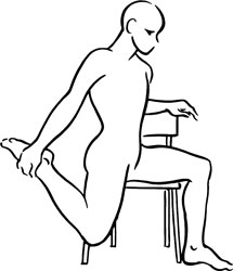
Stretch exercise 1:Tensor fasciae latae
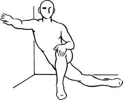
Stretch exercise 2:Tensor fasciae latae
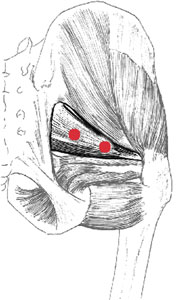
Piriformis and trigger points
P
IRIFORMIS
Proximal attachment:
Anterior surface of the sacrum.
Distal attachment:
Through the sciatic foramen, attaching to the greater trochanter of the femur.
Action:
External rotation of the thigh; acts in abduction when the thigh is flexed to 90 degrees.
Palpation:
To locate piriformis, identify the following structures:
- Greater trochanterâBony prominence on the lateral aspect of the femur, approximately one hand-length below the iliac crest. From the anterior plane the greater trochanter lies horizontal with the pubic crest.
- Piriformis lineâAn imaginary line drawn from the second sacral segment (just medial to the posterior superior iliac spine [PSIS]) to the upper border of the greater trochanter. This line represents the superior border of the piriformis muscle and the posterior border of the gluteus medius muscle.
Palpate piriformis with the patient side-lying or prone. Image the piriformis line; palpate slightly distal to that line, since it marks the superior border of the piriformis muscle. Palpate the muscle throughout its course, from the border of the sacrum to the greater trochanter. Taut bands of a constricted piriformis muscle can be palpated through gluteus maximus. Areas of constriction are most likely to develop in the medial aspect of the lateral one-third of the piriformis line and the lateral aspect of the medial one-third of that line.

Piriformis pain pattern
Pain pattern:
Pain in the sacroiliac region, the buttock, the posterior aspect of the hip joint, and possibly the proximal two-thirds of the posterior thigh. Pain is increased by sitting, standing, and walking.
Causative or perpetuating factors:
Acute overload; sustained overload due to immobilization in the externally rotated position; arthritis of the hip joint; pelvic inflammatory disease.
Satellite trigger points:
Gluteus medius, gluteus minimus.
Affected organ systems:
Genitourinary system; elimination aspect of the digestive system.
Associated zones, meridians, and points:
Dorsal and lateral zones; Foot Tai Yang Bladder meridian, Foot Shao Yang Gall Bladder meridian; GB 30.