Trigger Point Therapy for Myofascial Pain (26 page)
Read Trigger Point Therapy for Myofascial Pain Online
Authors: L.M.T. L.Ac. Donna Finando

Repeat the set (left/right) three to five times, increasing repetitions as strength allows.
For strengthening the abdominals as a group, you can also do the following two exercises.
- Roll downs: Begin in the seated position with the legs extended in front of you. Bend the knees, placing the soles of the feet on the floor. Inhale. Exhale, dropping the chin to the chest, rounding the lumbar spine, and slowly rolling backward toward the floor. First the lumbar spine touches the floor, then the thoracic spine, then the upper back, then the head. If need be you can control the backward movement of your weight by holding on to your legs as you roll back. Greater stress will be placed on the abdominal muscles the more slowly you roll down. Reverse the exercise to roll up.
- Abdominal crunches: Lying in the supine position bend the knees, placing the soles of the feet on the floor. Clasp the hands behind the head or cross the arms on the chest. Inhale deeply. Exhale, focusing on touching the navel to the spine, and roll up by bringing the chin to chest, raising the upper body, and lifting the scapulae off the floor. Slowly roll down to the starting position. Repeat three to five times, increasing repetitions as strength allows.
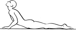
Stretch exercise 1: Abdominals
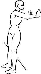
Stretch exercise 2: Abdominals
Â

Gluteus maximus and trigger points
G
LUTEUS
M
AXIMUS
Proximal attachment:
Posterior iliac crest, lateral sacrum, and coccyx.
Distal attachment:
Iliotibial band of the fasciae latae and the gluteal tuberosity of the femur.
Action:
Powerful extension of the thigh at the hip during strenuous activities such as running, jumping, stair climbing, and in rising from a seated position; helps maintain an erect posture; assists in lateral rotation of the hip.
Upper fibers:
abduction of the thigh.
Lower fibers:
adduction of the thigh.
Palpation:
To locate gluteus maximus, identify the following structures:
- Posterior superior iliac spine (PSIS)âBony prominence lying deep to the characteristic dimples above the buttocks. The PSIS lie horizontal to the second sacral segment. Each is approximately 2 centimeters (
 ) below the superior border of the sacrum.
 - Iliac crestâLying on a horizontal line with the junction of L4âL5.
- Sacrum
- Coccyx
- Greater trochanterâBony prominence on the lateral aspect of the femur, approximately one hand-length below the iliac crest. From the anterior plane the greater trochanter lies horizontal with the pubic crest.
- Ischial tuberosityâEasily palpable when seated, this bony prominence carries most of the weight of the torso in the seated position. It is located at the center of the buttock, approximately level with the gluteal fold.
To locate gluteus maximus, approximate its borders as follows: image its superior border by visualizing a line drawn from the PSIS to slightly above the greater trochanter; image its inferior border by visualizing a line drawn from the coccyx to the ischial tuberosity. To palpate gluteus maximus, follow the direction of muscle fibers obliquely and laterally, moving from the lateral margin of the sacrum to the greater trochanter.
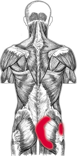
Gluteus maximus pain pattern
Pain pattern:
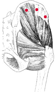
Medial trigger points, located adjacent to the sacrum, refer pain beside the gluteal cleft, including the sacroiliac joint. Distal trigger points, located above the ischial tuberosity, refer pain throughout the buttock, including tenderness deep within the buttock. Symptoms include local pain from prolonged sitting and increased pain when walking uphill in a forward-leaning position.
Causative or perpetuating factors:
Stress overload or impact from trauma or fall; prolonged walking in a forward-leaning position; injection.
Satellite trigger points:
Posterior gluteus medius, posterior gluteus minimus, hamstrings, iliopsoas, rectus femoris.
Affected organ system:
Elimination aspect of the digestive system; colon.
Associated zones, meridians, and points:
Dorsal and lateral zones; Foot Tai Yang Bladder meridian, Foot Shao Yang Gall Bladder meridian; BL 26â30, 35 and 36, 53 and 54, GB 30.
Stretch exercise:
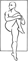
Lying supine, draw the knee toward the homolateral shoulder, grasping the posterior thigh; pull the thigh and leg toward the shoulder, stretching the gluteus maximus. Hold for a count of ten to fifteen. Release. Then draw the knee toward the opposite shoulder. Hold for a count of ten to fifteen and release.
Strengthening exercise:
Posterior pulses with a bent knee: Flex the leg of the affected side. Contract the gluteus maximus to extend the thigh. Repeat ten to twelve times.
Patient positioning will determine the degree of stress placed on the muscle.
Phase I (patient in the weakest condition):
Instruct the patient to do this exercise in the standing position, supporting his balance by holding onto a wall or chair.
Phase II:
Patient can be positioned side-lying, working the gluteal muscle of the upper leg.
Phase III:
Patient can be positioned on his hands and knees. Pulses will be working against gravity and will require the most force.

Stretch exercise: Gluteus maximus
(knee to homolateral shoulder)
Stretch exercise: Gluteus maximus
(knee to opposite shoulder)
Gluteus medius and trigger points