Trigger Point Therapy for Myofascial Pain (31 page)
Read Trigger Point Therapy for Myofascial Pain Online
Authors: L.M.T. L.Ac. Donna Finando

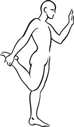
Stretch exercise:
Stand, or sit on the edge of a chair. Flex the lower leg and grasp the ankle with the homolateral hand. Lift the heel toward the buttock and extend the thigh and hip as far backward as possible. Tilt the pelvis to avoid excessive arching of the lumbar spine. Hold this position for a count of ten to fifteen.
Strengthening exercise:
Sit on a chair with the feet on the floor. Fully extend the working leg to a count of two. Return the leg to the starting position to a count of four. Repeat this exercise ten to twelve times, working one leg at a time.
Ankle weights may be used to increase the demand placed on the quadriceps during this exercise. The amount of weight used must be gauged according to the needs and capabilities of the patient.

Stretch exercise: Quadriceps

Adductor magnus

Adductor longus and Adductor brevis
Adductors and trigger points
A
DDUCTORS
A
DDUCTOR
M
AGNUS
, A
DDUCTOR
L
ONGUS
, A
DDUCTOR
B
REVIS
Proximal attachment:
Adductor magnus:
inferior pubic ramus, ramus of ischium, ischial tuberosity.
Adductor longus:
pubic tubercle.
Adductor brevis:
inferior ramus of the pubis.
Distal attachment:
Adductor magnus:
linea aspera of the posterior femur, adductor tubercle of the medial femur.
Adductor longus:
linea aspera on the middle one-third of the femur.
Adductor brevis:
proximal aspect of the linea aspera of the femur, lateral and deep to adductor longus.
Action:
Adductor magnus:
anterior fibers adduct and internally rotate the thigh; posterior fibers extend the thigh.
Adductor longus and adductor brevis:
adduction of the thigh at the hip.
Palpation:
Of the muscles in the adductor group, adductor longus is the most prominent and most readily accessible to palpation.
To locate the adductors, identify the following structures:
- Femoral triangleâBounded superiorly by the inguinal ligament, medially by adductor longus, and laterally by sartorius. The floor of the femoral triangle is formed medially by pectinius and laterally by iliopsoas. Within this triangle the femoral pulse can be palpated 2 to 3 centimeters (approximately 1 inch) inferior to the inguinal ligament, at the midline of the base of the triangle which it forms. Both the femoral artery and femoral lymph glands lie superficial to iliopsoas and pectinius, which themselves lie superficial to the hip joint. The femoral artery lies just superficial to the head of the femur. The pulse point of the femoral artery can be palpated just superficial to the head of the femur, inferior to the midpoint of the inguinal ligament.
- Ischial tuberosityâEasily palpable when seated, this bony prominence carries most of the weight of the torso in the seated position. It is located at the center of the buttock, approximately level with the gluteal fold.
Palpate adductor magnus and adductor longus with the patient lying supine. To palpate adductor magnus, flex the leg of the thigh to be palpated, then externally rotate and abduct the thigh in order to position the sole of the foot of that leg approximately 8 to 10 inches lateral to the inner thigh of the extended leg. If necessary, a pillow may be placed beneath the bent knee for patient comfort. Palpate adductor magnus posterior to adductor longus and adductor brevis, from the ischial tuberosity to the medial aspect of the femur.
To palpate adductor longus, place the sole of the foot against the inner thigh of the extended leg. In this position adductor longus becomes clearly visible and palpable. Palpate adductor longus anterior to adductor magnus, from its proximal aspect near the pubis to its distal aspect at the middle one-third of the femur.
Adductor longus and pectineus lie superficial to adductor brevis. Therefore adductor brevis cannot be directly palpated.

Adductor magnus

Adductor longus and Adductor brevis
Adductors pain pattern
Pain pattern:
Adductor magnus:
Trigger points in the proximal aspect of the muscle, near the ischial tuberosity, refer severe, deep pelvic pain that may include pain at the pubic bone, vagina, rectum, and possibly the bladder. Trigger points in the middle portion of adductor magnus refer pain to the anteromedial aspect of the thigh, from the groin to just above the knee.
Adductor longus and adductor brevis:
Pain in the groin that is experienced during activity; reduced abduction and external rotation of the thigh. Pain deep in the groin and possibly the anteromedial aspect of the upper thigh; pain above the medial aspect of the knee and possibly over the shin. Trigger points near the proximal attachment of adductor longus may cause knee pain and stiffness.
Causative or perpetuating factors:
Sudden overload due to a misstep or fall; arthritis of the hip joint; sustained overload due to activities such as horseback riding or sitting with the legs crossed for lengthy periods of time; emotional stresses.
Satellite trigger points:
Each muscle of the adductor group may develop satellite trigger points in response to the presence of trigger points in any other muscle of the group. Additional satellite trigger points could also appear in vastus medialis and gracilis.
Affected organ system:
Reproductive system.
Associated zones, meridians, and points:
Adductor magnus:
Ventral zone; Foot Jue Yin Liver meridian, Foot Shao Yin Kidney meridian; LIV 10 and 11, BL 36 and 37.
Adductor longus and adductor brevis:
ventral zone, Foot Jue Yin Liver meridian; LIV 9, 10, and 11.
Stretch exercises:
Adductor magnus forms a functional hamstring with the short head of the biceps femoris. Therapeutic stretching of the adductor group must therefore coincide with therapeutic stretching of the hamstring group in order to obtain the full benefit for each muscle group.
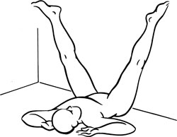
- Lying with the buttocks against a wall and the legs extended upward on the wall, slowly separate the legs to stretch the inner thighs. Hold this position for 30 to 60 seconds, allowing gravity to act on the abducted legs.
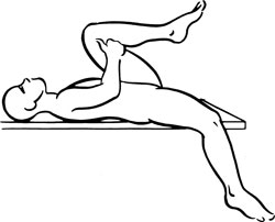
- Positioned supine, with the thigh and leg of the affected side off the side of an examining table or bed, flex the thigh and leg of the unaffected side to fix the pelvis, keeping the lumbar spine flat on the table. Abduct the thigh and leg, allowing it to hang off the side of the table or bed. The force of gravity will stretch the upper groin region. Hold this position for a count of 20 to 30.
Strengthening exercise:
Sitting on the edge of a chair, place a large, soft ball between the thighs. Adduct the thighs, squeezing the ball. Hold for a count of five to eight and release. Repeat ten to twelve times.

Stretch exercise 1: Adductors

Stretch exercise 2: Adductors

Pectineus and trigger point