Trigger Point Therapy for Myofascial Pain (32 page)
Read Trigger Point Therapy for Myofascial Pain Online
Authors: L.M.T. L.Ac. Donna Finando

P
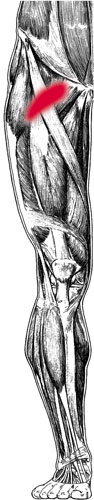
ECTINEUS
Proximal attachment:
Crest of the superior ramus of the pubis.
Distal attachment:
On the femur, just distal to the lesser trochanter.
Action:
Flexion, adduction, and internal rotation of the thigh at the hip.
Palpation:
To locate pectineus, identify the following structures:
- Femoral triangleâBounded superiorly by the inguinal ligament, medially by adductor longus (see muscle description on page 181) and laterally by sartorius (see muscle description on page 191). The floor of the femoral triangle is formed medially by pectineus and laterally by iliopsoas. Within this triangle the femoral pulse can be palpated 2 to 3 centimeters (approximately 1 inch) inferior to the inguinal ligament, at the midline of the base of the triangle that it forms. Both the femoral artery and femoral lymph glands lie superficial to iliopsoas and pectinius, which themselves lie superficial to the hip joint. The femoral artery lies just superficial to the head of the femur. The pulse point of the femoral artery can be palpatated just superficial to the head of the femur, inferior to the midpoint of the inguinal ligament.
- Pubic tubercleâThe bony prominence at the lateral aspect of the pubic crest. The pubic tubercle serves as the distal attachment for the inguinal ligament.
Palpate pectineus with the patient lying supine. Abduct and externally rotate the thigh to be palpated, then flex the leg to bring the sole of the foot adjacent to the opposite inner thigh. If necessary a pillow may be placed beneath the bent knee for patient comfort. Locate the femoral triangle and pubic tubercle. Pectineus occupies the medial aspect of the floor of the femoral triangle. To locate pectineus palpate within the femoral triangle, approximately 1 inch lateral and 1 inch distal to the pubic tubercle.
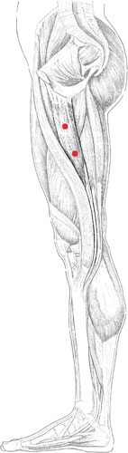
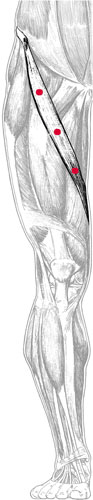
Pectineus pain pattern
Pain pattern:
Deep, local groin pain, just distal to the inguinal ligament.
Causative or perpetuating factors:
Sudden overload from an unexpected fall; chronic overload in adduction and flexion, such as sitting cross-legged for an extended period of time; diseases or surgeries affecting the hip joint.
Satellite trigger points:
Adductor longus, adductor brevis, adductor magnus, iliopsoas.
Affected organ system:
Reproductive system.
Associated zones, meridians, and points:
Ventral zone; Foot Jue Yin Liver meridian; LIV 9, 10, and 11.
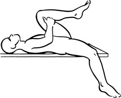
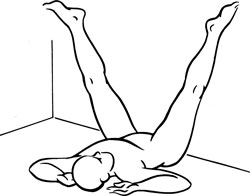
Stretch exercise:
Lying on a table or bed, abduct the affected thigh and leg and allow the limb to hang off the side of the table or bed. Flex the unaffected thigh and leg to fix the pelvis, keeping the lumbar spine flat on the table or bed. Let gravity stretch the upper groin region. Hold for a count of twenty to thirty.
Strengthening exercise:
Sitting on the edge of a chair, place a large, soft ball between the thighs. Adduct the thighs, squeezing the ball. Hold for a count of five to eight, and release. Repeat ten to twelve times.
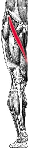
Stretch exercise: Pectineus
Gracilis and trigger points
Gracilis pain pattern
G
RACILIS
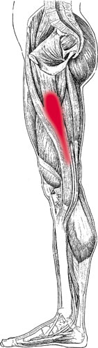
Proximal attachment:
Inferior pubic ramus.
Distal attachment:
Medial side of the superior part of the tibia, distal to the tibial condyle, forming the pes anserinus with sartorius and semitendinosus.
Action:
Adduction of the thigh at the hip; flexion of the leg; assists in internal rotation of the thigh and flexed leg.
Palpation:
To locate gracilis, identify the following structures:
- Medial surface of the tibial condyle
- Distal attachment of semitendinosusâIdentify the popliteal fossa, the posterior aspect of the knee joint that appears as a hollow when the knee is flexed. The distal attachment of semitendinosus forms the medial border of the popliteal fossa; the lateral border is formed by the tendon of biceps femoris.
Palpate the knee of the flexed leg at the popliteal region and identify the prominent tendon of semitendinosus forming the medial border of the popliteal fossa. Move slightly medially to locate the tendon of gracilis, which lies anterior and medial to the tendon of semitendinosus. Offering resistance to internal rotation of the thigh allows gracilis to become more prominent. Palpate this thin, bandlike muscle along its course on the medial aspect of the thigh.
Pain pattern:
Hot, stinging, superficial pain along the medial thigh.
Causative or perpetuating factors:
Sudden overload due to a misstep or fall; arthritis of the hip joint; sustained overload due to activities such as horseback riding or sitting with the legs crossed for lengthy periods of time; emotional stresses.
Satellite trigger points:
Sartorius.
Affected organ systems:
Reproductive and genitourinary systems.
Associated zones, meridians, and points:
Ventral zone; Foot Jue Yin Liver meridian; SP 9, LIV 8â11.
Stretch exercise:
Lying with the buttocks against a wall and the legs extended upward on the wall, slowly separate the legs to stretch the inner thighs. Hold this position for 30 to 60 seconds, allowing gravity to act on the abducted legs.
Strengthening exercise:
Sitting on the edge of a chair, place a large, soft ball between the thighs. Adduct the thighs, squeezing the ball. Hold for a count of five to eight, and release. Repeat ten to twelve times.
Stretch exercise: Gracilis
Sartorius and trigger points
Sartorius pain pattern
S
ARTORIUS
Proximal attachment:
Anterior superior iliac spine (ASIS).
Distal attachment:
Medial side of the superior part of the tibia, distal to the tibial condyle, forming the pes anserinus with semitendinosus and gracilis.
Action:
Assists in flexion, abduction, and external rotation of the thigh; assists in hip flexion and knee flexion during walking.
Palpation:
To locate sartorius, identify the following structures:
- Anterior superior iliac spine (ASIS)âAnterior bony projection lying somewhat below the iliac crest, readily palpable. The ASIS serves as the proximal attachment of the inguinal ligament.
- Medial surface of the tibial condyle
- Distal attachment of semitendinosusâIdentify the popliteal fossa, the posterior aspect of the knee joint that appears as a hollow when the knee is flexed. The distal attachment of semitendinosus forms the medial border of the popliteal fossa; the lateral border is formed by the tendon of biceps femoris.
Locate sartorius with the patient seated and the leg flexed to 90 degrees. Offering resistance to external rotation of the thigh, sartorius will become prominent from its attachment at the ASIS. Palpate sartorius from the ASIS throughout its course to its distal attachment, distal to the medial surface of the tibial condyle. The tendon of insertion of sartorius lies anterior to the tendons of insertion of gracilis and semitendinosus and forms the flattest and most anterior tendon of the pes anserinus.
Pain pattern:
Superficial tingling pain along the course of the muscle.