Trigger Point Therapy for Myofascial Pain (16 page)
Read Trigger Point Therapy for Myofascial Pain Online
Authors: L.M.T. L.Ac. Donna Finando

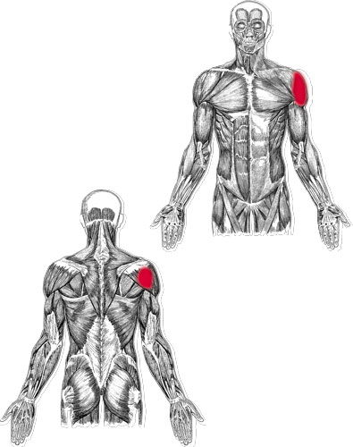
Deltoid (posterior) and trigger points
D
ELTOID
Proximal attachment:
Anterior fibers:
lateral one-third of the clavicle.
Medial fibers:
acromion.
Posterior fibers:
spine of the scapula.
Distal attachment:
Deltoid tuberosity of the humerus.
Action:
Anterior fibers:
flexion and internal rotation of the humerus.
Medial fibers:
abduction of the humerus.
Posterior fibers:
extension and external rotation of the humerus.
Palpation:
To locate the deltoid, identify the following structures:
- ClavicleâFollow the curved course of the clavicle, from its articulation with the sternum to its articulation with the acromion. Medially the contours of the clavicle are convex; laterally its contours are concave. The anterior deltoid attaches to the clavicle at its lateral concavity where the pectoralis major muscle ends.
- AcromionâThe flat, lateral aspect of the scapula at the most lateral tip of the shoulder girdle. Abducting the humerus as you palpate the lateral tip of the shoulder girdle, you can clearly distinguish between the acromion and the head of the humerus.
- Spine of the scapulaâFollow the course of the acromion posteriorly and medially along the spine of the scapula to the root of the spine of the scapula, a flattened, triangular surface at the medial border of the scapula. The root of the spine of the scapula is located on a horizontal line with the spinous process of T3.
- Deltoid tuberosity of the humerusâThe bony prominence located approximately midway down the lateral aspect of the humerus.
Palpate the deltoid from its attachments on the shoulder girdle to its attachment on the humerus. Palpate anteriorly, noting the area where the deltoid lies adjacent to the pectoralis at the lateral concavity of the clavicle. Note the deltopectoral groove formed by the junction of the deltoid and pectoralis major muscles. Palpate the medial fibers, noting the attachment of the deltoid muscle to the acromion; palpate posteriorly, noting the attachment to the spine of the scapula. Note how the three aspects of the muscle converge to insert onto the deltoid tuberosity of the humerus.
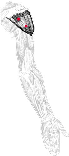
Deltoid pain pattern
Pain pattern:
Anterior fibers:
Pain is experienced in the anterior and medial deltoid; the patient may experience weakened abduction of the externally rotated arm.
Posterior fibers:
Pain is experienced in the posterior and medial deltoid; the patient may experience weakened abduction of the internally rotated arm.
Causative or perpetuating factors:
Direct trauma due to impact, overexertion, or sudden overload.
Satellite trigger points:
Anterior fibers:
pectoralis major, biceps brachii, posterior deltoid.
Posterior fibers:
long head of the triceps, latissimus dorsi, teres major.
Affected organ systems:
Anterior fibers:
respiratory system.
Posterior fibers:
digestive system.
Associated zones, meridians, and points:
Anterior fibers:
ventral zone; Hand Tai Yin Lung meridian; LU 1, 2, and 3.
Posterior fibers:
dorsal and lateral zones; Hand Yang Ming Colon meridian, Hand Shao Yang Triple Warmer meridian; CO 14 and 15, SI 10; TW 13 and 14.
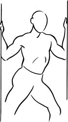
Stretch exercises:
- Anterior fibers
: Place the palms firmly on each side of a doorway at approximately ear level. Stretch the body through the outstretched arms, opening the chest and anterior shoulder region. - Posterior fibers:
Pull the affected arm across the chest, using the other arm placed proximal to the elbow to guide the action.
Strengthening exercises:
Both strengthening exercises should be performed in a standing position with the arms at the sides.
- Flex the affected armsâkeeping the elbows straight, bring the arm to shoulder level. Perform this flexion to a count of two; return to the starting position to a count of four. Repeat eight to ten times, increasing the number of repetitions as strength allows. Hand weights may be used to increase the work effort of the muscle.
- Abduct the affected arm, keeping the elbows straight; bring the arm to shoulder level. Abduct to a count of two; return to the starting position to a count of four. Repeat eight to ten times, increasing the number of repetitions as strength allows. Hand weights may be used to increase the work effort of the muscle.
Stretch exercise 1: Anterior deltoid

Stretch exercise 2: Posterior deltoid
Latissimus dorsi and trigger points
L
ATISSIMUS
D
ORSI
Proximal attachment:
Latissimus dorsi fuses with teres major to attach at the medial edge of the bicipital groove, on the anterior aspect of the humerus.
Distal attachment:
Spinous processes of T6âT12, L1âL5 and the sacrum, iliac crest via the thoracolumbar aponeurosis, lower 3â4 ribs, inferior angle of the scapula.
Action:
Extension, adduction, and internal rotation of the arm at the shoulder.
Palpation:
The latissimus dorsi and the teres major together form the posterior wall of the axilla. The tendon of the latissimus dorsi twists upon itself before fusing with teres major at the attachment on the bicipital groove.
To locate latissimus dorsi, identify the following structures:
- Spinous processes of T6 through T12 and L1 through L5âNote the difference in the size and shape of the spinous processes of the thoracic vertebrae versus the lumbar vertebrae.
- Iliac crestsâLying on a horizontal line with the junction of L4âL5.
- Inferior angle of the scapulaâThe sharp, triangular, distal aspect of the scapula. In most cases the inferior angle of the scapula lies on a horizontal line with T7.
- Bicipital groove of the humerus (intertubercular groove)âIdentify the greater and lesser tuberosities of the humerus, just distal to the lateral aspect of the acromion. (These are best palpated with the arm externally rotated.) The bicipital groove lies medial to the greater tuberosity and lateral to the lesser tuberosity. Note that the tendon of the long head of the biceps brachii runs through the bicipital groove.
Palpate latissimus dorsi with the patient in the prone position, arms resting at his sides, palms up. Use a pincer grasp at the posterior axillary fold, gently lifting the muscle off the thoracic wall. Grasp the muscle along its lateral placement, proximal to the inferior angle of the scapula, at approximately the midpoint of the lateral edge of the scapula; follow the muscle toward the iliac crest, where the muscle fibers become increasingly indistinct.
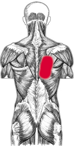
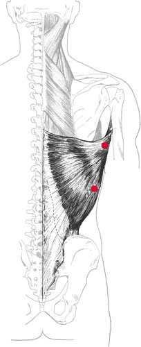
Latissimus dorsi pain pattern
Pain pattern:
Pain is located at the inferior angle of the scapula and the surrounding midthoracic region, possibly extending to the back of the shoulder and down the medial aspect of the arm, forearm, and hand, including the ring and little fingers. The nature of the pain is an ache that shows neither aggravation nor relief with activity or change of position.
Causative or perpetuating factors:
Depressor movements that overload, as in pulling something down from above or holding a heavy, bulky object.
Satellite trigger points:
Teres major, triceps brachii, rectus abdominis, iliocostalis thoracis, and iliocostalis lumborum.
Affected organ system:
Digestive system.
Associated zones, meridians, and points:
Dorsal and lateral zones; Foot Tai Yang Bladder meridian, Hand Tai Yang Small Intestine meridian; BL 23, SI 9.
Stretch exercise:
Reach both arms above the head. Grasp the wrist of the hand on the affected side with the opposite hand. Pull the wrist and arm toward the unaffected side, bending the torso to that side. Hold this position for a count of ten to fifteen.
Strengthening exercise:
Standing with the legs shoulder-width apart, bend from the waist so that the upper body is parallel to the floor. Reach the arm of the affected side toward the opposite foot. Bend the elbow to make a 90-degree angle and retract the scapula, extending the upper arm, bringing it to a position alongside the torso. The final position is one in which both the torso and the upper arm are parallel to the floor while the forearm is perpendicular to the floor. Draw the arm to the side to a count of two. Reach for the opposite foot to a count of four.