Trigger Point Therapy for Myofascial Pain (9 page)
Read Trigger Point Therapy for Myofascial Pain Online
Authors: L.M.T. L.Ac. Donna Finando

Stretch exercise 1: Clavicular head
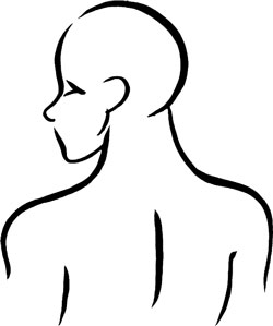
Stretch exercise 2: Sternal head

Scalenes and trigger points
S
CALENES
Proximal attachment:
Transverse processes of C2âC7.
Distal attachment:
Anterior and medius:
first rib.
Posterior:
second rib.
Action:
Acting unilaterally:
lateral flexion of the cervical spine.
Acting bilaterally:
stabilization of the cervical spine against lateral movement; elevation of the first and second ribs to assist in inspiration.
Palpation:
To locate the scalenes, identify the following structures:
- Thyroid cartilageâLocated along the anterior midline of the neck. The superior border of the thyroid cartilage, the Adam's apple, lies anterior to C4; the distal border of the thyroid cartilage lies anterior to C5. The thyroid cartilage can be observed moving up and down during swallowing.
- Hyoid boneâLocated above the thyroid cartilage and lying horizontally. The hyoid bone is the first bony structure to be palpated as you move downward along the midline from the mandible. It can be palpated as it moves up and down during swallowing. The hyoid bone lies anterior to C3.
- External jugular veinâBegins near the angle of the mandible, crossing the sterno cleidomastoid in the superficial fascia before passing posterior to the posterior border of the muscle, to empty into the subclavian vein. In the healthy patient, lying supine, the vein is clearly visible only a short distance above the clavicle. However, with increased thoracic pressure (noted in pathologies such as heart failure, enlarged supraclavicular lymph nodes, and obstruction of the superior vena cava) the external jugular vein becomes prominent throughout its course.
- Sternocleidomastoid muscleâSee muscle description on page 33.
The scalenes can be palpated when they are constricted or harbor trigger points. Note the location of the external jugular vein where it crosses the sternocleidomastoid: constriction of the anterior scalene can be palpated just deep to this area. Move the palpating finger slightly posteriorly to this area to locate the scalenes; they will feel like taut bands if they are constricted.
It is essential to palpate this area with the greatest sensitivity to patient tenderness and discomfort. An extremely gentle touch is required to be able to palpate the muscle without causing pain to the patient.

Scalenes pain pattern
Pain pattern:
Persistent, aching pain that radiates anteriorly and downward toward the chest in fingerlike projections and/or laterally to the upper arm. Pain may skip the elbow and reappear at the radial side of the forearm, hand, thumb, and index finger. Pain may radiate posteriorly into the midscapular area.
Causative or perpetuating factors:
Paradoxical breathing; chronic cough; pulling or lifting, especially with the arms level with the waist; chronic rotation of the cervical spine to one side.
Satellite trigger points:
Sternocleidomastoid, upper trapezius, levator scapula, splenius capitis, pectoralis major, triceps.
Affected organ system:
Respiratory system.
Associated zones, meridians, and points:
Ventral zone; Foot Yang Ming Stomach meridian; SI 16.
Stretch exercise:
Laterally bend the head and neck so that the ear of the unaffected side moves toward the same shoulder. Hold for a count of ten. Then, without changing the degree of lateral stretch, rotate the head and face toward the affected side, stretching the cheek toward the ceiling. Hold for a count of ten.
Return the head and face to the initial lateral stretch position. Now rotate the head and face, this time aiming the chin in toward the clavicle. Hold for a count of ten. Return the head and face to the initial lateral stretch position.
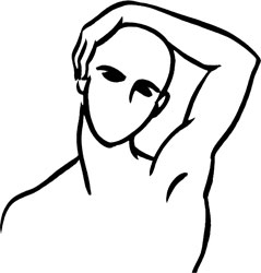
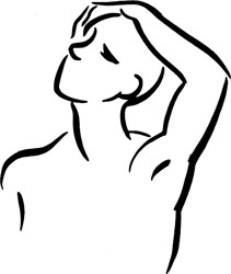
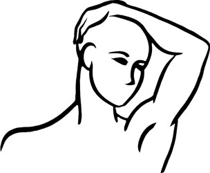
Stretch exercise: Scalenes
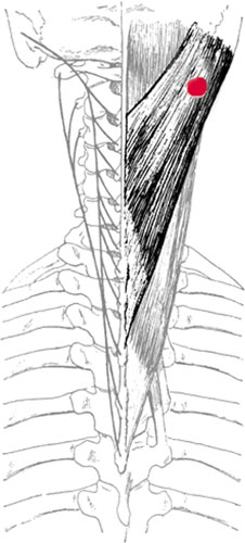
Splenius capitis and trigger point
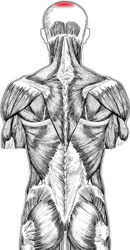
Splenius capitis pain pattern
S
PLENIUS
C
APITIS
Proximal attachment:
Mastoid process and adjacent occipital bone, deep to the attachment of the sternocleidomastoid muscle.
Distal attachment:
Fascia in the midline over the spinous processes of C4âT4.
Action:
Acting unilaterally:
rotation of the head and neck to the same side.
Acting bilaterally:
extension of the head and neck.
Palpation:
To locate splenius capitis, identify the following structures:
- SternocleidomastoidâSee muscle description on page 33.
- TrapeziusâSee muscle description on page 59.
- Levator scapulaeâSee muscle description on page 63.
To palpate splenius capitis, place your patient in the seated position with his back resting on the back of the chair, or lying supine. Palpate the portion of splenius capitis that lies just above the angle of the neck: locate the muscular triangle that is formed by the sternocleidomastoid muscle (anteriorly), the trapezius muscle (posteriorly), and the levator scapulae (distally). Gently palpate taut bands of splenius capitis just proximal to the levator scapulae.
Pain pattern:
Pain located at the vertex of the head.
Causative or perpetuating factors:
Postural stresses that overload, such as thrusting the head forward to compensate for excessive thoracic kyphosis.
Satellite trigger points:
Levator scapulae, upper trapezius, ste ocleidomastoid, splenius cervicis.
Affected organ system:
Vision.
Associated zones, meridians, and points:
Dorsal zone; Foot Tai Yang Bladder meridian.
Stretch exercise:
Rotate the head 20 to 30 degrees toward the unaffected side. Gently press the head forward and toward the unaffected side, stretching slightly more forward than laterally.
Strengthening exercise:
Isometric against mild posterior resistance. Clasp the hands behind the head at the level of the nuchal line. Press the head and neck posteriorly against the mild resistance provided by the clasped hands.

Stretch exercise: Splenius capitis