The Singularity Is Near: When Humans Transcend Biology (28 page)
Read The Singularity Is Near: When Humans Transcend Biology Online
Authors: Ray Kurzweil
Tags: #Non-Fiction, #Fringe Science, #Retail, #Technology, #Amazon.com

Peering into the Brain
We’ve been able to reduce drift and noise in our instruments to such an extent that we can see the tiniest motions of these molecules, through distances that are less than their own diameters. . . . [T]hese kinds of experiments were just pipedreams 15 years ago.
—S
TEVEN
B
LOCK, PROFESSOR OF BIOLOGICAL SCIENCES AND OF APPLIED PHYSICS
, S
TANFORD
U
NIVERSITY
Imagine that we were trying to reverse engineer a computer without knowing anything about it (the “black box” approach). We might start by placing arrays of magnetic sensors around the device. We would notice that during operations that updated a database, significant activity was taking place in a particular circuit board. We would be likely to take note that there was also action in the hard disk during these operations. (Indeed, listening to the hard disk has always been one crude window into what a computer is doing.)
We might then theorize that the disk had something to do with the longterm
memory that stores the databases and that the circuit board that is active during these operations was involved in transforming the data to be stored. This tells us approximately where and when the operations are taking place but relatively little about how these tasks are accomplished.
If the computer’s registers (temporary memory locations) were connected to front-panel lights (as was the case with early computers), we would see certain patterns of light flickering that indicated rapid changes in the states of these registers during periods when the computer was analyzing data but relatively slow changes when the computer was transmitting data. We might then theorize that these lights reflected changes in logic state during some kind of analytic behavior. Such insights would be accurate but crude and would fail to provide us with a theory of operation or any insights as to how information is actually coded or transformed.
The hypothetical situation described above mirrors the sort of efforts that have been undertaken to scan and model the human brain with the crude tools that have historically been available. Most models based on contemporary brain-scanning research (utilizing such methods as fMRI, MEG, and others discussed below) are only suggestive of the underlying mechanisms. Although these studies are valuable, their crude spatial and temporal resolution is not adequate for reverse engineering the salient features of the brain.
New Tools for Scanning the Brain
. Now imagine, in our computer example above, that we are able to actually place precise sensors at specific points in the circuitry and that these sensors are capable of tracking specific signals at very high speeds. We would now have the tools needed to follow the actual information being transformed in real time, and we would be able to create a detailed description of how the circuits actually work. This is, in fact, exactly how electrical engineers go about understanding and debugging circuits such as computer boards (to reverse engineer a competitor’s product, for example), using logic analyzers that visualize computer signals.
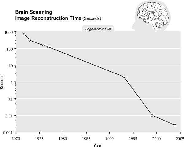
Neuroscience has not yet had access to sensor technology that would achieve this type of analysis, but that situation is about to change. Our tools for peering into our brains are improving at an exponential pace. The resolution of noninvasive brain-scanning devices is doubling about every twelve months (per unit volume).
31

We see comparable improvements in the speed of brain scanning image reconstruction:
The most commonly used brain-scanning tool is fMRI, which provides relatively high spatial resolution of one to three millimeters (not high enough to image individual neurons) but low temporal (time) resolution of a few seconds. Recent generations of fMRI technology provide time resolution of about one second, or a tenth of a second for a thin brain slice.
Another commonly used technique is MEG, which measures weak magnetic fields outside the skull, produced principally by the pyramidal neurons of the cortex. MEG is capable of rapid (one millisecond) temporal resolution but only very crude spatial resolution, about one centimeter.
Fritz Sommer, a principal investigator at Redwood Neuroscience Institute, is developing methods of combining fMRI and MEG to improve the spatiotemporal precision of the measurements. Other recent advances have demonstrated fMRI techniques capable of mapping regions called columnar and laminar structures, which are only a fraction of a millimeter wide, and of detecting tasks that take place in tens of milliseconds.
32
fMRI and a related scanning technique using positrons called positronemission
tomography (PET) both gauge neuronal activity through indirect means. PET measures regional cerebral blood flow (rCBF), while fMRI measures blood-oxygen levels.
33
Although the relationship of these blood-flow amounts to neural activity is the subject of some controversy, the consensus is that they reflect local synaptic activity, not the spiking of neurons. The relationship of neural activity to blood flow was first articulated in the late nineteenth century.
34
A limitation of fMRI, however, is that the relationship of blood flow to synaptic activity is not direct: a variety of metabolic mechanisms affect the relationship between the two phenomena.
However, both PET and fMRI are believed to be most reliable for measuring relative changes in brain state. The primary method they use is the “subtraction paradigm,” which can show regions that are most active during particular tasks.
35
This procedure involves subtracting data produced by a scan when the subject is
not
performing an activity from data produced while the subject
is
performing a specified mental activity. The difference represents the change in brain state.
An invasive technique that provides high spatial and temporal resolution is “optical imaging,” which involves removing part of the skull, staining the living brain tissue with a dye that fluoresces upon neural activity, and then imaging the emitted light with a digital camera. Since optical imaging requires surgery, it has been used mainly in animal, particularly mouse, experiments.
Another approach to identifying brain functionality in different regions is transcranial magnetic stimulation (TMS), which involves applying a strongpulsed magnetic field from outside the skull, using a magnetic coil precisely positioned over the head. By either stimulating or inducing a “virtual lesion” of (by temporarily disabling) small regions of the brain, skills can be diminished or enhanced.
36
TMS can also be used to study the relationship of different areas of the brain on specific tasks and can even induce sensations of mystical experiences.
37
Brain scientist Allan Snyder has reported that about 40 percent of his test subjects hooked up to TMS display significant new skills, many of which are remarkable, such as drawing abilities.
38
If we have the option of destroying the brain that we are scanning, dramatically higher spatial resolution becomes possible. Scanning a frozen brain is feasible today, though not yet at sufficient speed or bandwidth to fully map all interconnections. But again, in accordance with the law of accelerating returns, this potential is growing exponentially, as are all other facets of brain scanning.
Carnegie Mellon University’s Andreas Nowatzyk is scanning the nervous system of the brain and body of a mouse with a resolution of less than two hundred nanometers, which is approaching the resolution needed for full reverse engineering. Another destructive scanner called the “Brain Tissue Scanner”
developed at the Brain Networks Laboratory at Texas A&M University is able to scan an entire mouse brain at a resolution of 250 nanometers in one month, using slices.
39
Improving Resolution
. Many new brain-scanning technologies now in development are dramatically improving both temporal and spatial resolution. This new generation of sensing and scanning systems is providing the tools needed to develop models with unprecedented fine levels of detail. Following is a small sample of these emerging imaging and sensing systems.
One particularly exciting new scanning camera is being developed at the University of Pennsylvania Neuroengineering Research Laboratory, led by Leif H. Finkel.
40
The optical system’s projected spatial resolution will be high enough to image individual neurons and at one-millisecond time resolution, which is sufficient to record the firing of each neuron.
Initial versions are able to scan about one hundred cells simultaneously, at a depth of up to ten microns from the camera. A future version will image up to one thousand simultaneous cells, at a distance of up to 150 microns from the camera and at submillisecond time resolution. The system can scan neural tissue in vivo (in a living brain) while an animal is engaged in a mental task, although the brain surface must be exposed. The neural tissue is stained to generate voltage-dependent fluorescence, which is picked up by the highresolution camera. The scanning system will be used to examine the brains of animals before and after they learn specific perceptual skills. This system combines the fast (one millisecond) temporary resolution of MEG while being able to image individual neurons and connections.
Methods have also been developed to noninvasively activate neurons or even a specific part of a neuron in a temporally and spatially precise manner. One approach, involving photons, uses a direct “two-photon” excitation, called “two-photon laser scanning microscopy” (TPLSM).
41
This creates a single point of focus in three-dimensional space that allows very high-resolution scanning. It utilizes laser pulses lasting only one millionth of one billionth of a second (10
−15
second) to detect the excitation of single synapses in the intact brain by measuring the intracellular calcium accumulation associated with the activation of synaptic receptors.
42
Although the method destroys an insignificant amount of tissue, it provides extremely high-resolution images of individual dendritic spines and synapses in action.
This technique has been used to perform ultraprecise intracellular surgery. Physicist Eric Mazur and his colleagues at Harvard University have demonstrated its ability to execute precise modifications of cells, such as severing an
interneuronal connection or destroying a single mitochondrion (the cell’s energy source) without affecting other cellular components. “It generates the heat of the sun,” says Mazur’s colleague Donald Ingber, “but only for quintillionths of a second, and in a very small space.”
Another technique, called “multielectrode recording,” uses an array of electrodes to record simultaneously the activity of a large number of neurons with very high (submillisecond) temporal resolution.
43
Also, a noninvasive technique called second-harmonic generation (SHG) microscopy is able “to study cells in action,” explains lead developer Daniel Dombeck, a graduate student at Cornell University. Yet another technique, called optical coherence imaging (OCI), uses coherent light (lightwaves that are all aligned in the same phase) to create holographic three-dimensional images of cell clusters.
Scanning Using Nanobots
. Although these largely noninvasive means of scanning the brain from outside the skull are rapidly improving, the most powerful approach to capturing every salient neural detail will be to scan it from inside. By the 2020s nanobot technology will be viable, and brain scanning will be one of its prominent applications. As described earlier nanobots are robots that will be the size of human blood cells (seven to eight microns) or even smaller.
44
Billions of them could travel through every brain capillary, scanning each relevant neural feature from up close. Using high-speed wireless communication, the nanobots would communicate with one another and with computers compiling the brain-scan database. (In other words, the nanobots and computers will all be on a wireless local area network.)
45