Social: Why Our Brains Are Wired to Connect (23 page)
Read Social: Why Our Brains Are Wired to Connect Online
Authors: Matthew D. Lieberman
Tags: #Psychology, #Social Psychology, #Science, #Life Sciences, #Neuroscience, #Neuropsychology

BOOK: Social: Why Our Brains Are Wired to Connect
2.84Mb size Format: txt, pdf, ePub
There’s a second limitation to the near exclusive focus on empathy for pain within neuroimaging research.
The neural systems responsible for affect matching should vary as a function of what kind of affect is being matched.
Given that nearly all of the follow-ups to Singer’s seminal work have focused on empathy
for physical pain, one could easily review this literature and come to the conclusion that the dACC and the anterior insula are the central mechanisms supporting empathy in general.
Is this really the case, or are these regions showing up because they are involved in pain and most of the studies focus on pain?
Finally,
almost none of the studies that have been done have linked neural responses
during the empathic state to actual helping behavior.
One of the purposes of feeling empathy seems to be to motivate us to help others in distress, yet it’s unclear how the brain converts our understanding and affect matching processes into the empathic motivation to help.
Sylvia Morelli, Lian Rameson, and I ran an fMRI study
that we hoped would capture all three components of empathy: understanding, affect matching, and empathic motivation.
First, we varied whether context was needed in order for the key event to be understood.
Some of the time, we showed participants a picture of someone experiencing pain that showed all that they needed to know to immediately grasp the event (for example, a hand being slammed in a car door).
On other trials, the picture showed someone
looking happy or anxious and required context to be understood (for example, “This person is waiting to get his medical test results”).
The first kind of event recruited the mirror system; the context-dependent events recruited the mentalizing system.
In addition, though the happy and anxious events both included context, they required different kinds of affect matching because they involved different kinds of emotional events.
The anxious events and the pain events both activated the pain distress network, but the happy events instead activated a region of the ventromedial pre-frontal cortex that is commonly recruited during reward tasks.
Perhaps most important, we searched for brain regions that were commonly activated across all three kinds of empathy events we had included (pain, anxiety, and happiness).
Our thinking was that while understanding and affect matching differ depending on the content driving one’s empathy, the ultimate empathic motivation to help should be the end result in each case.
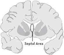
There was only one brain region that was activated during each type of empathy event: the septal area (see
Figure 7.1
).
In addition to showing up in each condition, the septal area appeared to be a marker of empathic motivation.
The subjects that we put in the scanner filled out a survey each day for two weeks about things they did and did not experience each day.
Among the questions we asked each day was whether they had helped someone else during that day.
By averaging across the two weeks of responses, we had a measure of which people tended to help others more frequently in their daily life.
Those who showed more activity in the septal area when they performed our empathy task in the scanner were the same people who tended to help other people more often outside the scanner.
This is consistent with the notion that the septal area takes the converging inputs from other brain regions involved in empathy and converts them to the urge to be helpful.
What was different on the night that I donated to help a child in Africa, in contrast to the countless other times when I changed the channel?
Probably septal area activity.

Figure 7.1 The Septal Area
The Septal Area
If I had to bet on which brain region is most ignored by the field of social neuroscience today but will be
the hot area of study in the next ten years
, the septal area would be it.
This structure has become disproportionately larger across primate evolution, and it has direct connections to
the dorsomedial prefrontal cortex (DMPFC)—the CEO of the brain’s mentalizing system
.
The vast majority of the work on the septal area has been done in rodents, rather than humans, in part because it is such a tiny region that it is hard to identify with fMRI.
The downside to studying rodents is that we can’t measure their experiences or even verify that they have them.
The upside is that more invasive studies can be conducted to examine how individual septal neurons respond or how surgical removal of the septal area alters behavior.
Animal research provides clues to what the septal area does, but these clues seem to lead us down different paths.
Some of the earliest research on the septal area focused on pleasure and reward.
Although the ventral striatum has far more often been identified with reward, its neural neighbor, the septal area, was actually the first brain region identified with reward processes.
The brain’s reward system was first discovered in the 1950s when electrodes were
implanted into various brain regions of rats and hooked up to a lever.
When a rat pressed the lever
, one of the brain regions was electrically stimulated.
When the electrodes were placed in the septal area, the rats went wild.
One rat pressed the lever nearly 2,000 times per hour—more than once every two seconds, nonstop for an hour.
Two decades later a similar study was conducted
with a man who had electrodes implanted in three locations and was given a button box with a button to stimulate each of the three regions.
Just like the rats before him, he pressed the button for the septal region relentlessly, indicating that it gave him intense pleasure, and he complained at the end of each session when the button box was taken from him.
At the same time that researchers were linking the septal area to reward
, other researchers were showing its involvement in fear or, more accurately, its role in reducing fear behavior.
One of the best measures of anxiety or fearfulness is called the
startle response.
If someone were to clap her hands loudly behind your head unexpectedly, there would be a cascade of neural, physiological, and behavioral responses that would code this noise as a potential threat and prepare you to respond quickly—a classic fight or flight response.
You would probably jump up, turn around, and perhaps notice that your heart was racing.
These responses are orchestrated by the amygdala, a phylogenetically ancient structure in the brain often associated with emotional responding.
Rats whose septal area has been removed show a much larger startle response and show other evidence of being more reactive to threats.
This suggests that when the septal area is intact, it may function to dampen the distress we feel in response to threats.
Last but not least, a separate body of research suggests the septal area is critical for maternal caregiving.
Lesion studies in rats, mice, and rabbits suggest
that if the septal area is damaged, the animal will be a terrible parent.
These lesioned animals no longer make protective nests for their young, they provide their young with less milk, and they experience a much higher rate of infant mortality.
How do we make sense of the various functions of the septal area—reward, fear regulation, and maternal caregiving?
Recent work by Tristen Inagaki and Naomi Eisenberger suggests that
one way to reconcile the findings is to characterize the septal area as shifting the balance
between our approach and avoidance motivations, which promotes proactive parenting.
Although humans start planning for their baby’s arrival months or even years in advance, most mammals probably do not have the same kind of logical understanding of their relationship to their newborn infants.
In the absence of this knowledge in most mammals, screaming babies are a real dilemma.
Should we rush to help them or run for the hills?
Mammals are wired to fear noisy uncertain things, but the septal area may help to quiet our fears and increase our motivation to help those in need.
Instead of selfishly taking cover, we selflessly put ourselves in the line of fire.
Thus, the septal region appears to be the key node that converts our affective responses into the motivation to provide help.
It is no accident that this description of the septal area parallels the
nurse neuropeptide
account of oxytocin we saw in the context of social rewards.
The septal area is rich in oxytocin receptors
, and, for some mammals, this region has the highest density of oxytocin receptors in the brain.
Intriguingly, this density is affected by early parenting experiences.
Among rodents, pups who receive more parental care
grow up to have higher oxytocin receptor density in the septal area, whereas pups who are separated from their mothers grow up to have lower oxytocin receptor density in the septal area.
Empathy is arguably the pinnacle of our social cognitive achievements—the peak of the social brain.
It requires us to understand the inner emotional worlds of other people and then act in ways that benefit other people and our relationships with them.
It can motivate us to alleviate another’s pain or to celebrate someone else’s good fortune.
All of the neural mechanisms that we have talked about so far need to be coordinated in order to make this amazing achievement possible.
Depending on the situation, we
need the mirror and/or mentalizing systems to understand someone else’s experience.
We need the mechanisms that support social pains and pleasures for the affect matching that allows us to feel, not just know, the other’s experience.
Finally, we need the septal region, central to maternal caregiving, to nudge us to actually get involved in the lives of those around us in positive ways.
When all of these mechanisms are in place, we can be our best selves.
Being a Social Alien
In 1992, the year I graduated from Rutgers, I had one of the worst days of my life.
As someone who had a long-standing interest in the mind, how it constructs reality, and in science fiction authors like Philip K.
Dick who are famous for creating alternate realities, it was more or less inevitable that I would dabble in mind-altering substances as a young adult.
If the mind was flexible and reality flexed with it, how could I pass up a firsthand demonstration?
There’s little point in identifying the particular drug I took that afternoon because the truth was that you never quite knew what you were getting.
My roommates from 12 Prosper Street and I headed over to a midday celebration at Cook Campus a few weeks before graduation at Rutgers.
We all took the same drug on the way over, something we had all taken before.
Everyone had a great time but me.
I had the proverbial “bad trip.”
There was nothing I could do but ride it out.
I have replayed this day in my head so many times, if only to remind myself why drugs are not my friend.
In all the times I have revisited this horror film, I never once thought about how I must have seemed to those around me.
They couldn’t tell what I was experiencing internally.
Most had no idea I had ingested anything more powerful than bad keg beer, and those who did know were too busy having fun to care much.
I must have come across as very odd, awkward, and antisocial (which, hopefully, is not how I come across the rest of the time).
I kept my distance from people, kept
my answers very short, and avoided eye contact.
I wonder if I didn’t seem a bit autistic that day.
Autism is a profound disorder affecting nearly 1 percent of the population.
Its dominant symptoms include repetitive behaviors and impairments in social interaction and verbal communication.
Asperger’s syndrome shares the difficulties in social interaction without the additional language deficits.
Clinically speaking, current diagnoses are for
autism spectrum disorders
(ASDs).
If empathy is the peak of the social mind, autism is sadly one of its low points.
There have been many theories over the years as to why these individuals have so much difficulty with the social world.
As we will see, things are sometimes almost the exact opposite of how they seem.
Just two years after the first Sally-Anne test for Theory of Mind
was performed, British psychologists Simon Baron-Cohen (yes, Sasha’s cousin), Alan Leslie, and Uta Frith proposed that ASD individuals may lack a Theory of Mind.
Can you imagine a world in which you didn’t see other people’s actions in terms of their beliefs, goals, and feelings?
Try doing it for a few minutes in your next social encounter.
You probably can’t, which only shows how much a part of our basic operating system this capacity is.
But if you could, it would make you feel a bit like an alien—the bodies in motion around you meaning nothing more than the surface features that you saw.
Actions would seem random and unpredictable without your being able to “see” the mind behind them.
Could you hold down a job, have friends, or maintain a long-term relationship seeing the world through that lens?
Not having a Theory of Mind would seem to explain many of the difficulties individuals with autism have in daily life.
Other books
Waco's Badge by J. T. Edson
Darkening Skies by Bronwyn Parry
Switch by William Bayer
Great Soul: Mahatma Gandhi and His Struggle With India by Joseph Lelyveld
Eric S. Brown by Last Stand in a Dead Land
Stormswept by Sabrina Jeffries
Time Present and Time Past by Deirdre Madden
To Protect An Heiress (Zebra Historical Romance) by Basso, Adrienne
A Brighter Fear by Kerry Drewery