In The Blink Of An Eye (32 page)
Read In The Blink Of An Eye Online
Authors: Andrew Parker

Compound eyes


The post-Cambrian view

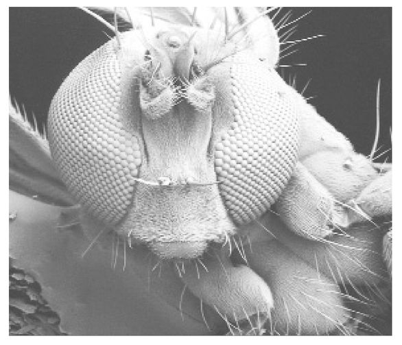
Figure 7.4
Scanning electron micrograph of the head of a fly, showing compound eyes.
Scanning electron micrograph of the head of a fly, showing compound eyes.
By way of introduction to compound eyes, I will return briefly to the somewhat unrefined ocelli. There is one group of bristle worms with ocelli but these are different from those of other animals. The difference lies in their arrangement. Lying on thick, feather-like filaments sprouting from the head, the ocelli of these worms are grouped together.
Each ocellus has a sac-like region formed as an outgrowth of a sensory hair. This region lies within an infold in the skin of the animal and acts as a lens. Behind this lies a well-developed region of light-sensitive chemicals - the âretina'. And within a group of ocelli, light-absorbing pigment cells intermingle to prevent the same light rays affecting more than one ocellus. But the information collected by each ocellus is later combined strategically and so elaborate composite organs are formed. An organ of this type is known as a compound eye (although these particular eyes fall a little short of the visual mark).
In contrast to the simple eye, the compound eye has multiple openings for light to enter - hence its name - and so always consists of numerous individual units, or ocelli, called âfacets'. Other than minor appearances in the bristle worms and ark clams, the compound eye is a character of the arthropods. More precisely, compound eyes today occur in crustaceans, insects and horseshoe âcrabs' (which are actually more closely related to scorpions than true crabs). Compound eyes have evolved into sophisticated organs of sight, up to a third of the total body size in some seed-shrimps, and form images in different ways.
The law of compound eyes was laid in 1891 in a monograph by the biologist Sigmund Exner, which became a landmark to both biologists and optical theorists. Exner broke all the rules of his day, where simple eye concepts were being applied to compound eyes. Instead Exner considered the focusing elements of compound eyes as âlens cylinders'. A conventional lens relies on the bending of light as it crosses a curved surface to focus rays. A lens cylinder, on the other hand, gently persuades light to change direction throughout the cylinder's length. It is literally a cylinder, but one filled with graded material - graded in terms of its effect on light, just like the fish lens contemplated by Maxwell. The lens cylinder is most dense, and so causes light to travel slowest, along its central axis, its ability to bend light fading towards the edges. The overall effect of a lens cylinder is to provide the image-forming properties of a traditional lens. But there are alternatives to lens cylinders in some compound eyes.
The compound eyes of many insects and crustaceans have a similar superficial appearance, but their focusing elements and mechanisms of
image formation are very different. We can divide compound eyes into two basic types - apposition and superposition. The facets of apposition eyes are optically isolated from each other, so they each sample a different section of the environment. The tiny images formed within each facet are pieced together in the manner of a jigsaw puzzle to produce the complete picture. The facets of superposition eyes, on the other hand, cooperate optically so that they superimpose their light to form a single image at a common point on the retina. Dividing compound eyes further, there are variations of both apposition and superposition compound eyes in terms of focusing and image formation.
image formation are very different. We can divide compound eyes into two basic types - apposition and superposition. The facets of apposition eyes are optically isolated from each other, so they each sample a different section of the environment. The tiny images formed within each facet are pieced together in the manner of a jigsaw puzzle to produce the complete picture. The facets of superposition eyes, on the other hand, cooperate optically so that they superimpose their light to form a single image at a common point on the retina. Dividing compound eyes further, there are variations of both apposition and superposition compound eyes in terms of focusing and image formation.

Figure 7.5
Focusing mechanisms in the compound eyes of a) bees (apposition-type eye); b) moths and c) lobsters (superposition-type eyes). Graded material in b) and mirrors in c) (shown from the side and from above) achieve focusing. (Modified from Land, 1981.)
Focusing mechanisms in the compound eyes of a) bees (apposition-type eye); b) moths and c) lobsters (superposition-type eyes). Graded material in b) and mirrors in c) (shown from the side and from above) achieve focusing. (Modified from Land, 1981.)
Superposition eyes contain a large number of facets - up to several hundred. A broad zone of clear material separates the lenses from the retina below - the facets are not continuous tubes, so light can cross from the lens of one facet to the receptor of another. Exner discovered this after considering the cornea of the glow-worm (actually a beetle) as a single structure rather than as individual units. The cornea forms the lenses of the glow-worm, and Exner simply cleared out the interior of a glow-worm eye so that he could examine the complete array of lenses. Instead of a series of inverted images corresponding with each lens, he found just one upright image. So all the lenses must focus on to the same point on the retina.
Some crustacean eyes are without lens cylinders, and it was not until 1975 that an alternative focusing mechanism was discovered. While examining the eyes of a crayfish and a deep-sea shrimp respectively, Michael Land and the German biologist Klaus Vogt independently found a superposition eye in which each facet was lined with mirrors. The mirrors were again similar to those found in fish skin, and formed mirror boxes, square in section. Exner got as far as illustrating the shape of these boxes, but overlooked the silver inner surfaces. It is now clear that if the boxes are considered with mirrored sides, the light rays will change direction as they are reflected and will all meet at the same point - on the retina where they form an image. In other words, mirror boxes perform the task of focusing.
In 1988 a third form of superposition eye was discovered in many crabs by another contemporary expert on eyes - Dan-Eric Nilsson of Lund University in Sweden. The optics in this case are complex, and involve an intricate combination of ordinary lenses, cylindrical lenses, parabolic mirrors and light guides. The imaging mechanism is equally elaborate, with three separate systems in operation. Image formation can be predicted from the hardware alone, suggesting that fossil eyes could be informative after all.
Compound eyes have no iris to control light levels, but they do have an alternative solution - they use dark pigment to remove some light where needed. This is similar to the way cats and crocodiles use pigment, but the pigment is in a different part of the eye. When superposition eyes are exposed to high light levels, the dark pigment
moves between the lenses and retina to absorb a proportion of the light rays. When light levels become extreme, sometimes the facets become optically separated by dense collars or tubes of absorbing pigment so that the eye effectively acts as an apposition eye.
moves between the lenses and retina to absorb a proportion of the light rays. When light levels become extreme, sometimes the facets become optically separated by dense collars or tubes of absorbing pigment so that the eye effectively acts as an apposition eye.
All in all, the architecture or optics of modern eyes are well understood and can teach us much about the way their hosts see. And architecture can be preserved in the remains of extinct animals. We are now well enough informed to be able to browse the library on fossil eyes.
Ancestral eyesThe post-Cambrian view
Conodonts are animals named after the Greek word meaning âcone teeth'. This is because for some time they were known only from jaw-like structures and bone fragments. Conodonts evolved in the Cambrian and became extinct 220 million years ago. They have been used extensively for comparing and assessing the age of rock sequences, but until the early 1980s we had no idea what the conodont animal looked like. Then complete fossil conodonts, around 340 million years old, were found in the Granton Shrimp Beds near Edinburgh, Scotland. These fossils revealed animals of eel-like appearance, with tails containing supporting fin rays . . . and heads with large, camera-type eyes.
The smaller species of conodonts have eyes which are larger in relation to their body length than are the eyes of the larger species of conodonts in relation to theirs. This is consistent with a rule of the relative growth of the eye dating back to 1762, which holds that smaller animals have larger eyes in relation to their body size than do larger animals. From work on living vertebrates, eye size is known to influence visual sharpness. And the fact that conodonts possessed reasonably large eyes carries important information on early vertebrate evolution.
Theories that conodonts are larval stages of âagnathans', the primitive jawless fishes that include today's lampreys and hagfishes, have not been well received. Agnathans are the first representatives of the true
vertebrates, within the chordate phylum, which evolved around 485 million years ago, just after the Cambrian. But the relatively small eyes and large bodies of some conodonts are evidence against their interpretation as juvenile agnathans. So conodonts are more generally believed to be chordates close to, but not part of, the line
leading
to true vertebrates. The eyes tipped the balance of opinion in this direction.
Among the living agnathans, only hagfishes have âeyes' that are smaller than those of conodonts. But the âeyes' of hagfishes, those primitive fishes caught in the deeper-water SEAS traps, are rather light perceivers - they do not yield visual images. They have probably degenerated as an adaptation to dark environments and burrowing. On the other hand, the eyes of lampreys are well developed and generally larger than those of conodonts. But there is one group of lampreys - the smallest brook lampreys - which offers clues to the vision of conodonts.
Small brook lampreys have eyes that are about a millimetre and a half in diameter, equal in size to the eyes of the conodont
Clydagnathus
. There is evidence to suggest that similarity in the size of camera-type eyes reflects a similarity in cell and nervous complexity - the information processing system. The finding of eye muscles in another, well-preserved conodont,
Promissum
, supports this line of thinking - the muscles of similar-sized eyes are also equivalent.
The conclusions drawn from a comparison with small brook lampreys are that conodonts had pattern vision and, as will become relevant later in this book, an active predatory lifestyle. Nevertheless, to say that smaller conodonts had relatively larger eyes does not imply that vision played a greater role in their behaviour. Instead they possessed visual organs near the minimum size limit for âeyes' - these organs could not have produced visual images if they were smaller.
Thorius
, a miniaturised salamander which is the smallest land vertebrate living today, has camera-type eyes that are little over a millimetre in diameter, and this is believed to be the lowest size limit that will provide precise vision.
Thorius
, a miniaturised salamander which is the smallest land vertebrate living today, has camera-type eyes that are little over a millimetre in diameter, and this is believed to be the lowest size limit that will provide precise vision.
At the larger practical limit of eye size was
Ophthalmosaurus
, a dolphin-shaped reptile between 3 and 4 metres in length. While dinosaurs were evolving big bodies on land,
Ophthalmosaurus
was
setting a record for camera-type eyes in the sea. This animal really did possess eyes the size of soccer balls, and they were used to see at depths of 500 metres and more. It is thought that this reptile dived deep to avoid predators or to catch deep-dwelling prey. Unfortunately
Ophthalmosaurus
suffered from âthe bends', a condition familiar to deep-sea divers who approach the surface too rapidly. Rapid ascent causes nitrogen gas dissolved in the blood to decompress, forming bubbles that can block blood vessels and kill tissue. The bends leave visible depressions in the joints of bones, and those depressions are evident in the fossils of
Ophthalmosaurus
. The bends, and its effects, occurred much less commonly with its smaller-eyed ancestors, but the deep-diving, big-eyed version was stopped in its evolutionary tracks - and so the
Ophthalmosaurus
died out.
Ophthalmosaurus
, a dolphin-shaped reptile between 3 and 4 metres in length. While dinosaurs were evolving big bodies on land,
Ophthalmosaurus
was
setting a record for camera-type eyes in the sea. This animal really did possess eyes the size of soccer balls, and they were used to see at depths of 500 metres and more. It is thought that this reptile dived deep to avoid predators or to catch deep-dwelling prey. Unfortunately
Ophthalmosaurus
suffered from âthe bends', a condition familiar to deep-sea divers who approach the surface too rapidly. Rapid ascent causes nitrogen gas dissolved in the blood to decompress, forming bubbles that can block blood vessels and kill tissue. The bends leave visible depressions in the joints of bones, and those depressions are evident in the fossils of
Ophthalmosaurus
. The bends, and its effects, occurred much less commonly with its smaller-eyed ancestors, but the deep-diving, big-eyed version was stopped in its evolutionary tracks - and so the
Ophthalmosaurus
died out.
Remaining within the ancient vertebrates, early fossil fishes commonly have dark stains in the region of the head. But what do these stains represent? The eyes of one particular agnathan specimen can provide an answer. This fossil fish has, in the exact position of the stains found in other agnathans, an eyeball fully preserved in a sub-spherical hardened structure with a slit directed sideways. Alex Richie, an expert on primitive fishes at the Australian Museum, has interpreted the stains as the remains of pliable, presumably cartilaginous, structures surrounding the actual eyeball. The earliest of these camera-type eyes are known from a 430-million-year-old specimen of
Jamoytius kerwoodi
. Richie believes that although not specifically found in the eyes of this fossil specimen, there is no doubt that a lens of some form was present, based on comparisons with these camera-type eyes found in later, related species.
Jamoytius kerwoodi
. Richie believes that although not specifically found in the eyes of this fossil specimen, there is no doubt that a lens of some form was present, based on comparisons with these camera-type eyes found in later, related species.
Other books
Faithful in Pleasure by Lacey Thorn
Due or Die by Jenn McKinlay
Fixing Hell by Larry C. James, Gregory A. Freeman
The Real Prom Queens of Westfield High by Laurie Boyle Crompton
Infected by V.A. Brandon
Constricted: Beyond the Brothel Walls by Ryans, Rae
Confessions After Dark by Kahlen Aymes
Dark Universe by Devon Herrera
Wayward Hearts by Susan Anne Mason