In The Blink Of An Eye (29 page)
Read In The Blink Of An Eye Online
Authors: Andrew Parker

Structures such as the feathers of birds have been preserved at Messel as if they had only moments earlier fallen from the sky, but in my biased opinion the greatest treasures of all at Messel - and justification alone for the high security - are the metallic-coloured beetles. Their optical effects are extraordinary. Stag beetles reflect the shimmering blues and greens they displayed while alive. As the shale containing a jewel beetle is broken, the sight of 49-million-year-old iridescent yellows and reds is revealed. And so the list of beetles and colours continues.
Sometime in 1997 I received a parcel from Germany from the palaeontologist Stephan Schaal, a man whose name is almost synonymous with that of Messel. As I had hoped, it contained Messel beetles
from a recent excavation. The fossils were stored in water, and the wing cases shimmered with violet, blue and green. Since colour in animals was at the centre of my research, the first question to cross my mind was, âWhat is causing this colour?' The age of the fossils simply did not register - the beetles looked like zoological museum specimens recently collected from a rainforest expedition. After all, 49 million years is a long period to comprehend or time travel in one's mind.
from a recent excavation. The fossils were stored in water, and the wing cases shimmered with violet, blue and green. Since colour in animals was at the centre of my research, the first question to cross my mind was, âWhat is causing this colour?' The age of the fossils simply did not register - the beetles looked like zoological museum specimens recently collected from a rainforest expedition. After all, 49 million years is a long period to comprehend or time travel in one's mind.
To answer my question, I turned to electron microscopy. Small sections of a blue beetle exoskeleton were treated in two different ways. One section was critical point dried - that is, it was dried out in a controlled manner to prevent shrinkage. Although it had retained its structure, the dried section had lost its colour. It had become transparent. To examine the structure in the scanning electron microscope, it was first coated with gold. Then, at 10,000 times magnification, thin layers became evident, with upper layers only partly overlapping the lower layers. The layers were smooth, and there was no sign of a diffraction grating or structures that could cause the scattering of light. But to confirm that this was a multilayer reflector, transmission electron micrographs were needed.
One of the beetle sections was embedded in resin, stained and sliced so thin that it was not visible edge on. Placed on a minute metal grid to provide support, the specimen was imaged in an electron beam. A multilayer reflector was revealed.
To be doubly sure, the dimensions of the reflector were measured and fed into a computer program which re-created the stack of thin layers and predicted the colour of the light reflected in sunlight at 90° to the surface. The predicted colour was blue. The actual colour I saw was blue. Hence the cause of the colour of Messel beetles
was
a multilayer reflector. The reason the colour had disappeared from the dried specimen also could have been predicted. It emerged that one of the two layer types in the reflector consisted of water - when the water disappeared, so did the colour.
was
a multilayer reflector. The reason the colour had disappeared from the dried specimen also could have been predicted. It emerged that one of the two layer types in the reflector consisted of water - when the water disappeared, so did the colour.
Several specimens of the same beetle species have been found at Messel, and all display exactly the same colours. So we can be confident that 49 million years ago beetles were gracing Europe with spectacular iridescence - last seen flashing when the dead beetles were
washed into the Messel lake by floodwater, and were sinking into the depths of history. Re-opening the history book, we learn that light must have been a powerful stimulus to animal behaviour even then. But how far back can structural colours help us take this philosophy?
Fossils of the Burgess Shale - diffraction gratingswashed into the Messel lake by floodwater, and were sinking into the depths of history. Re-opening the history book, we learn that light must have been a powerful stimulus to animal behaviour even then. But how far back can structural colours help us take this philosophy?
In 1966 Kenneth Towe and Charles Harper, palaeontologists at the Smithsonian Institution, published a paper describing the cause of iridescence in 420-million-year-old lamp shells. They found tubular aragonite crystals arranged in layers, with dimensions in the region of the wavelength of light. A layer of juxtaposed tubes may create a diffraction grating on the outside, but a stack of thin layers can also form a multilayer reflector. The lamp shells appeared with a rather faint iridescence or pearly lustre like that of some shells today. Towe and Harper suggested the cause of this optical effect was a combined grating-multilayer structure, and attributed the faintness to variations in spacings, or a degree of randomness in the structure. Further work may be required to confirm these conclusions, but we cannot say for certain that these colours were sparkling in 420-million-year-old waters. Again, considering shells today, the lamp shell iridescence may have been precluded by an opaque outer layer, a layer that was not preserved in the fossil.
True diffraction gratings are well known to physicists, but before my search, prompted by their discovery in seed-shrimps, they were unknown in nature. Then diffraction gratings began to appear in one animal after another. First there was a lobster found off Hawaii, then a type of shrimp from New Caledonia, again in the Pacific Ocean. But the Indian Ocean was hiding similar treasures, not only within its crustaceans but in bristle worms, comb jellies, jellyfish and peanut worms. Eventually it was discovered that the entire globe contained a vast array of species, from many animal phyla, loaded with diffraction gratings. The world, it turned out, was even more colourful than we had believed it to be, albeit that the newly revealed iridescence was often concealed from view for most of the time.
Part of my work on seed-shrimp iridescence described in Chapter 5 was carried out at the National Museum of Natural History of the Smithsonian Institution. Originally I had found diffraction gratings in some seed-shrimps from Australia and needed to examine as many other species as possible. The world's expert on this group of animals is Louis Kornicker at the Smithsonian, and it is no coincidence that the best collection of seed-shrimps is found there, too. So it was only natural that I should apply for funding to work in Washington. My application was successful and in 1995 I began working on the Smithsonian collection.
As mentioned in Chapter 1, the Smithsonian also houses probably the best and certainly the most important collection of Burgess Shale fossils anywhere in the world. Now that is a coincidence. The Smithsonian was the home of Charles Doolittle Walcott, who discovered the first Burgess Shale fossils. But other than a general fascination in this âwonderful life' evident among all zoologists, I had no specific interest in the fossils themselves.
Taking a break from work at the Smithsonian, one is spoilt for choice for things to do. Within a few blocks of each other on a single avenue there are several national museums and art galleries. But there was also the Museum of Natural History, and during one late afternoon break I found myself wandering around the fossil galleries.
I discovered a small but excellent exhibit on the Burgess Shale nestling between larger skeletons. This exhibit was worthy of its space because the fossils displayed were complete and detailed examples of the range of life forms for which, in addition to its age, the Burgess Shale was famous. The specimens were also those collected mainly by Walcott in the early 1900s.
Next to each fossil in the exhibit were black and white illustrations showing reconstructions of the animals when they were alive. The drawings were very detailed and really helped one to visualise the
living
creatures. But the level of detail included something of interest to me specifically. On some reconstructions there was a hint of something quite amazing. On the reconstructed armoured parts of
Hallucigenia
and
Wiwaxia
were fine parallel lines. And fine parallel lines were the reason I had come to Washington in the first place.
The day before, I had visited the aviation and space museum which housed some aeroplanes from the 1950s, each with multiple propellers and corrugated wings and fuselage. The corrugations served to increase the strength of the metal structures. Later I was to encounter similar corrugations used to increase structural strength - but in the leaves of a Rocky Mountain plant on my expedition to the Burgess Shale quarry. These leaves were thin and would have collapsed were it not for their corrugated form. This was important to bear in mind. Narrow striations on the Burgess Shale fossils could represent a finely corrugated surface to make them stronger. But it got me thinking. The same rules apply to animals today, although if the striations meet certain size criteria, they cause iridescence - they become diffraction gratings.
Reference to diffraction gratings insinuates microscopically fine corrugations, where a distance approaching the wavelength of light separates neighbouring ridges. Such structures cannot be drawn as lines on paper. No pen is that sharp or precise, and we would not be able to see the lines with the naked eye anyway. But, as I have said, this got me thinking. Maybe the lines figured in the animal reconstructions were merely representatives of diffraction gratings. Fossil preservation is never uniform, and perhaps only some ridges of a grating had been preserved. Then again, maybe the lines figured were complete and did serve to provide strength. If this were the case, the parallel lines illustrated in the Smithsonian exhibit would add nothing to the topic of colour in fossils. But they changed the direction of my thoughts. If animals possess diffraction gratings today, maybe they did so in the past too.
The morning after my first Burgess Shale viewing, I submitted a request to examine the original Cambrian finds. Access to the Smithsonian fossils, and those at Harvard University, was granted, following support from Simon Conway Morris of Cambridge University, Doug Erwin at the Smithsonian and Frederick Collier at Harvard. Then there were some specimens to examine at the Australian Museum back in Sydney. To begin with I employed the most powerful light microscopes available at the Smithsonian Institution. I had not realised that the compact disc case I was using to orientate the specimens under the microscope was titled âHandel's Water Music' - quite appropriate, as certain onlookers remarked. But it worked. I placed the fossils so they
could be viewed from different angles, and structures I had not noticed before became evident. Then I knew exactly what to look at, and the work became serious. I took the fossils to the various underground rooms of different institutions, where vibrations and magnetic fields that could interfere with more powerful microscopes were minimal. And there the microscope cavalry charged in to the project. By the end of my experiments, I had bombarded many species of Burgess animals with a barrage of laser and electron beams, and had imaged the specimens at extremely high magnifications - so high that even single molecules could be observed.
could be viewed from different angles, and structures I had not noticed before became evident. Then I knew exactly what to look at, and the work became serious. I took the fossils to the various underground rooms of different institutions, where vibrations and magnetic fields that could interfere with more powerful microscopes were minimal. And there the microscope cavalry charged in to the project. By the end of my experiments, I had bombarded many species of Burgess animals with a barrage of laser and electron beams, and had imaged the specimens at extremely high magnifications - so high that even single molecules could be observed.
The techniques I used were all harmless to the fossils, which in some cases included original organic material, but there was one further test I wanted to carry out which would have altered the fossils permanently. The scanning electron microscope exacts a thin coat of metal to be applied to any animal surface under observation - a coat that cannot be removed practically. So rather than harming the invaluable fossils, casts were made. Plaster of Paris can be used to make casts of dinosaur footprints, but the Burgess Shale fossils under investigation were small and the diffraction gratings are microscopic. The particles in plaster of Paris are simply too large to fill the grooves of a diffraction grating and produce a detailed cast. But I had learnt of a new technique using acetate, and this enabled fine, elaborate casts to be made. When dry, the casts rather than the fossils were gold-coated and could be examined in a scanning electron microscope.
After the last microscopic tests had been completed, the potentially amazing became a reality. The reactions of several electron microscopy technicians indicated that the results were both positive and special. On the broken surfaces of three species - the bristle worms
Wiwaxia
and
Canadia
, and the arthropod
Marrella
- were remnants of diffraction gratings. Only traces of gratings had been preserved, rather like the few squares that remain in many Roman mosaics, but where they did occur on a single body part, they were always exactly the same size and shape, and were orientated in the same direction. The results were consistent. But the fragmentation had extinguished iridescence in the actual fossils. The fossils were decidedly grey. The mood in my lab, on the other hand, was more colourful.
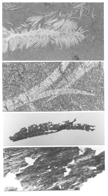
Figure 6.1
Micrographs of the Burgess bristle worm
Canadia
at increasing magnification - from
x
10 to
x
1,500. The top picture shows the front half of the animal, the middle pictures show details of bristles. The bottom picture shows the surface of a bristle as removed from the rock matrix, revealing the remnants of a diffraction grating with a ridge spacing of 0.9 microns.
Did this really mean that
Wiwaxia
,
Canadia
and
Marrella
would have appeared highly coloured when they lived 515 million years ago? This still seemed unbelievable. To make doubly sure, the original surfaces of
Canadia
and
Marrella
were reconstructed in their entirety, based on the remnants that had preserved. This was achieved by carefully positioning two laser beams so they met and interfered at the surface of a light-sensitive material and etched out the precise sinusoidal contours of the remnant gratings over the entire material (the model was examined further to confirm this). The reconstructed surfaces were taken out of the dark laboratory and placed in seawater under sunlight, and . . . the colours of three Burgess Shale species shone as spectacularly as they had 515 million years ago. That was the most memorable moment of all. For the first time, the original colour of a Cambrian animal had been uncovered. An almost unimaginable piece of Cambrian history had been revealed.
Other books
Tomorrow Happens by David Brin, Deb Geisler, James Burns
Love in Xxchange: Rory's Last Chance by Bailey Bradford
Wicked: The Original Broadway Script by Carter, Devin
The King's Wizard by James Mallory
Left Behind by Jayton Young
A Kingdom of Dreams by Judith McNaught
A Goal for Joaquin by Jerry McGinley