Planet of the Bugs: Evolution and the Rise of Insects (25 page)
Read Planet of the Bugs: Evolution and the Rise of Insects Online
Authors: Scott Richard Shaw

The transport of eggs, hypodermic-fashion, across a microscopically thin tubular pathway required the evolution of microscopic eggs
with highly flexible shells, eggs that could be distorted from an oval shape into a long, thin, sausage shape while being forced through this minute tubule. But more importantly, wasp eggs needed a mechanism that would transport them through the ovipositor. In this case, the mechanism was fluid pressure: the ovipositor is a hypodermic needle through which wasp eggs are literally squirted. Consequently, right from the start, wasp ovipositors evolved with a variety of associated liquids. These fluids initially came from female reproductive glands, and provided not only lubrication inside the ovipositor shaft, but also fluid pressure for physically transporting the eggs. They also allowed for the early evolution of wasp venoms.
From sawflies to wood wasps to parasitic wasps, these venoms diversified to accomplish an array of useful functions that we still see today. Some sawflies inject venoms into plant tissues along with eggs, and these venoms induce unusual plant cell development, causing galls to grow. These growths form protective sites for egg development, as well as safe areas and nutritious tissues for larval feeding. In other cases the injected venoms may have antibiotic properties that protect the eggs from the ravages of microbial growth. Among wood wasps, gooey venoms were adapted to promote the growth of fungus. As I mentioned in previous chapters, wood, with its high amounts of nonnutritive lignin and cellulose, is the least digestible part of the plant for an insect. So, like the wood-boring beetles before them, wood wasps adapted to eat the fungus that grows inside decaying wood.
The immature wood wasps were bound to bump into the juicy larvae of various wood-boring beetles from time to time, which were bound to be more delicious and nutritious than either rotting wood or fungi. The jump from chewing on fungi to chewing on the meat of other insects does not seem all that great when one considers that on the tree of earthly life, animals are more closely related to fungi than we are related to plants. In other words, animal tissue is more like the tissue of a mushroom than that of a leafy green plant. Nutritionally and physiologically, the change was not that enormous.
More challenging, perhaps, were the vital behavioral changes involved in switching from a vegetarian to a carnivorous life style. A wood wasp chewing on fungi in a rotting log didn’t have to contend with the fungi fighting back. Beetle grubs, however, were not totally defenseless. They could still wiggle around in their tunnels, and when
attacked they could bite just as well as the wasps. Moreover, a wood wasp’s developing eggs would be defenseless against a beetle larva’s chewing mouthparts. Clearly the wood wasps needed a secret weapon to tip the scales in their favor.

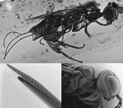
FIGURE 8.2. Megalyrid wasps (order Hymenoptera, family Megalyridae) are among the oldest living examples of parasitoid wasps; the family is thought to have evolved in the Jurassic period.
Top
, a female of an undescribed extinct megalyrid species in mid-Cretaceous amber from Myanmar with a long ovipositor, estimated to be about ninety-nine million years old. (Photo by Vincent Perrichot.)
Bottom
, the head (
right
) and ovipositor (
left
) of a modern, undescribed
Dinapsis
species from Madagascar, a rare surviving example of this ancient wasp group.
The adult wasps, not their larvae, developed the trick that gave them the decisive advantage, and once again, the female ovipositor made all the difference. At about the same time that some wood wasp larvae were developing a taste for meat, some of their mothers were refining their egg-laying skills and trying out new kinds of venoms. These mother wasps would drill adroitly into the tunnels where large beetle larvae were feeding, then poke their ovipositors directly into
the larvae and inject a new kind of venom that induced permanent paralysis. Then they would extract their ovipositor a bit and carefully place their eggs on the paralyzed beetle. Consider the advantage of venom that did not kill an insect, but rendered it motionless. If a beetle larva were killed by an adult wasp, it would begin to rot and decay, and the decomposition process would endanger the developing wasp egg. Paralyzing the host insect with venom instead is a cheap and efficient way to preserve the meat until the egg can hatch and the baby wasp can safely begin feeding. It’s pretty much like setting a full food dish next to a lazy dog: the wasp larva doesn’t have much to do other than sit there and chew on a big chunk of fresh meat.
Which Way to Eat an Oreo: Two Kinds of Parasitism
The sort of external parasitism that I’ve been describing has existed for about 150 million years. Termed “ectoparasitism,” which literally means “feeding as a parasite from the outside,” it became, over the intervening years, an important factor in the evolution and success of the wasps in particular. But the origin of parasitism is really just the beginning of a much bigger story. Although the first parasitoid species were very successful, diversifying and chewing on whatever young beetles they could find in the decaying wood for millions of years, they stayed restricted to that particular habitat until some entrepreneurial wasp came along with another new approach. Sometime in the Late Jurassic or Early Cretaceous the parasitoid wasps figured out how to feed inside other animals as an internal parasite. This more refined method of parasitism is what we term endoparasitism. It literally means “feeding as a parasite from the inside,” and looking at the modern insect world, the vast majority of parasitic species are of this second sort.
Although ectoparasitoids’ early success in the Middle Jurassic was mainly due to their strategy of feeding externally on only one small organism, this approach had a major drawback. The host insect was permanently paralyzed and the immature wasp needed ample time to hatch from its egg, then devour the entire beetle grub. The process took many weeks, at least, and could work only in concealment. If exposed, the young wasp would be revealed to predators and subject to harsher environmental factors: desiccating sunlight, temperature ex
tremes, wind, and storms. Therefore external parasitism was mostly limited to protected microhabitats inside plant tissue, which hindered what ectoparasitoids could ultimately achieve. Now, as then, the great majority of external-feeding parasitoids are associated with immature insects found inside plant tissues. The internal feeders, in contrast, were unfettered by these habitat constraints. The host became the endoparasitic wasp’s niche and its entire habitat during its youthful existence (endoparasitism was literally the discovery of niches within other insects). Once inside the host, the endoparasitic wasp larva became entirely portable, and it could exist in any habitat where the host might exist. As a result, endoparasitic wasps were able to diversify and feed inside virtually all kinds of Mesozoic insects. A Pandora’s box of feeding behaviors was opened for the wasps.
Once again, an important event in the history of wasps was the refinement of egg-laying behavior and the female ovipositor. Some females quit laying their eggs on the outside of the host and started injecting them directly inside it. Stated succinctly like that, the transition to endoparasitism sounds simple, but it wasn’t. Remember that female wasps were already drilling and stinging host insects for millions of years, injecting paralyzing venoms with their ovipositors. They could have easily inserted their eggs inside other insects along with venom as soon as they developed parasitic behavior. But they didn’t. Instead, they pulled out their ovipositors from inside the hosts and laid their eggs on the outside, allowing their young wasp larvae to feed externally. The reason they didn’t initially place their eggs on the inside is that being there is a lot more challenging than being on the outside. Insects have an open circulatory system, so their inside is a sack full of organs bathed in a pool of blood. That blood, like our own, contains cells that defend insects against microscopic invasion. Early parasitic wasp venoms may have paralyzed a host’s muscular system, but they did not incapacitate the immune response of its blood cells. A small egg placed inside an insect’s body cavity would be swarmed and encapsulated by these cells and killed. Successful endoparasitism required that wasps evolve an array of special adaptations to the internal environment.
The first step to successful internal parasitism was yet another refinement in precision egg laying: some wasps laid eggs directly into the host’s nerve or muscle tissue, thereby avoiding its blood—and
immune system—entirely. This is a nice adaptation, as far as it goes, but sooner or later the egg needs to hatch and the young larva must move around and eat. It’s hard to avoid the blood entirely, and in fact, there is a very good reason to want to be there: insect blood is a pool of nutrient-rich fluids. So ultimately the most successful internal parasitoids were the ones that invented ways to compromise the host’s immune system. Once again the mother wasps helped their offspring. Along with eggs, they injected venoms, some of which were modified to help disable the immune system—but somewhere along the line an even more unexpected event occurred: a symbiotic relationship was forged between certain viruses and the wasps.
We all have heard how dirty hypodermic needles can transfer viruses. Back in the very early days of internal parasitism, one of the wasps managed to soil its own hypodermic ovipositor with some virus particles. This happened fortuitously, but then those particles were injected, along with a wasp egg, into a hapless host insect. The virus replicated itself within the host, disabling its immune system but not harming the wasp larva. At the same time, the virus was able to imbed itself inside the developing wasp’s body and so was able to escape and eventually find its way to another potential host. It was a win-win situation for both the virus and the wasp.
7
Once host immune systems were disabled, wasp eggs and larvae could wallow safely in insect blood. An external parasite’s egg has a tough outer shell, which protects it from environmental factors, and a large protein-rich yolk, which feeds the developing embryo until it hatches into a larva. An egg placed in blood, on the other hand, does not have such a thick outer shell; it floats in a protein-rich liquid environment and, with a thin shell, absorbs nutrients directly from the host’s blood. Now the endoparasitic wasps only needed a mechanism for extracting nutrients, so that their eggs could survive with little, if any, yolk. If less yolk were needed, then females could produce eggs more easily and lay more of them. And so the endoparasitic wasps evolved a structure called a trophamnion, which works like a parasitic placenta. The trophamnion consists of a cluster of cells, closely connected to the embryo, that absorbs and transfers nutrients directly from the host’s blood. These cells feed the embryo it until it develops into a larva, which bursts from its egg and swims away. The trophamnion’s benefits do not end there. As the egg hatches, its cells disassoci
ate and move independently into the host’s blood pool. These former trophamnion cells continue to extract nutrients from the insect’s blood and work to further disable its immune system. As they continue to feed, they grow and eventually morph into giant cells, called teratocytes, which the young wasp larva also consumes as it swims about and dines on the host’s blood and tissues.
Wasp larvae adapted to life in their miniature aquatic environment by developing the ability to swim with long, taillike appendages, which they whip back and forth, and the ability to breathe with closed, gas-filled tracheal systems, which, since they have no open breathing holes, prevent water from flooding into them. The larvae also developed a thin cuticle without much hard skeletal material along their body wall. This allowed them to breathe directly through their body by a process known as cuticular respiration, just like many other aquatic insects that live in ponds and streams. Indeed, the wasps are actually the tiniest of aquatic insects and also the most diversified group of aquatic organisms.
Although a larger aquatic insect living in a pond has a lot of room to move about, a parasitic wasp larva swimming in insect blood is limited to a small enclosed space. It literally lives inside a bowl of soup, its only food. This presents a special problem: anything that eats and grows must also produce waste, so how does the larva continue to develop without fouling its food and living environment? We can appreciate the importance of teaching kids not to pee and poop in the swimming pool and bath tub—all the more important when the pool is a food source as well. Young wasps solve this problem by simply accumulating waste inside their bodies and never defecating. They assimilate nutrients very efficiently with the middle part of the digestive system, but the hind part is closed, forcing waste into the rear end. When a wasp larva is done feeding, growing, and molting through several stages, it exits the host to spin its own silk cocoon, pupate, and finally transform into an adult. Upon emerging from its cocoon, the full-grown wasp voids its larval waste for the first and last time.