Pediatric Examination and Board Review (187 page)
Read Pediatric Examination and Board Review Online
Authors: Robert Daum,Jason Canel

2.
(D)
Serology is the method of choice for diagnosis. Both IgG and IgM antibodies to
B henselae
can be measured. Most patients with catscratch disease have high IgG antibodies at presentation. The IgM test lacks sensitivity. If lymph node tissue is available, the organism may sometimes be seen with the Warthin-Starry silver impregnation stain, but this stain is not specific for
B henselae
.
3.
(B)
Surgical excision of lymph nodes is unnecessary. Antimicrobial therapy may be beneficial for severely ill patients with systemic catscratch disease and is recommended for immunocompromised patients. There are two other clinical syndromes of
B henselae
and
B quintana
infections reported in immunocompromised patients. Bacillary angiomatosis is a vascular proliferative disorder that involves the skin and subcutaneous tissues and occurs in immunocompromised individuals. Bacillary peliosis occurs primarily in patients with AIDS and is characterized by reticuloendothelial lesions in the liver primarily that can also involve the spleen, abdominal lymph nodes, and bone marrow. The lesions of these two diseases respond to treatment with erythromycin, doxycycline, or azithromycin.
4.
(E)
Treatment is recommended only for patients who have a history of, or active peptic ulcer disease, gastric mucosa-associated lymphoid tissue-type lymphoma (MALToma), or early gastric cancer. The most effective regimen in children includes a 2-week, 3-agent therapy that consists of a protein pump inhibitor such as omeprazole or lansoprazole, clarithromycin, and amoxicillin.
5.
(A)
The child likely has brucellosis with osteoarticular involvement. Childhood brucellosis most often affects the large peripheral joints, including the knees, hips, and ankles. A definitive diagnosis is established by recovery of
Brucella
species from blood, bone marrow, or other tissues. If brucellosis is suspected, the clinical microbiology laboratory personnel should be informed so blood cultures can be incubated for 4 weeks. A serum agglutination test (SAT) and EIA are also available for diagnosis. It is recommended to send the SAT first and measure antibody titers in serum specimens collected at least 2 weeks apart.
6.
(B)
The adolescent has a clinical form of tularemia called oculoglandular syndrome that results from conjunctival infection and is acquired from contaminated fingers.
F tularensis
can be transmitted by direct contact with infected animals, through tick bites, and also by contaminated food or water. The most common clinical manifestation is the ulceroglandular syndrome. This disease manifests as swollen, tender lymph nodes in the inguinal, cervical, or axillary regions that are preceded by painful maculopapular lesions at the portal of entry that develop into an ulcerated lesion (
Table 105-1
).
7.
(E)
Osteoarticular infection is the most common clinical infection caused by
K kingae
in children. Studies have shown that inoculating of joint fluid directly into BACTEC aerobic blood culture bottles increases the likelihood of isolating the bacteria. Most infections occur in children younger than 5 years of age.
K kingae
is a common cause of septic arthritis in young children in Israel but has been reported less frequently in the United States.
TABLE 105-1
Clinical Manifestations of Tularemia
| CLINICAL SYNDROME | COMMENT |
Ulceroglandular | Most common form; adenitis ± ulcer |
Oculoglandular | Nodular conjunctivitis; enlarged painful preauricular nodes |
Oropharyngeal | Pseudomembrane-simulating diphtheria; fever; associated with ingestion of contaminated meat, milk, or water |
Typhoidal tularemia | High fever; signs of sepsis; hepatosplenomegaly common; ingestion of contaminated food; can have necrotic lesions in bowel |
Gastrointestinal | Ingestion of contaminated food; persistent diarrhea and abdominal or back pain |
8.
(A)
The most important clinical infection caused by
L pneumophila
in both children and adults is pneumonia. Infections in immunocompromised children such as among those in receipt of anticancer therapy represent the most severe form of the disease in pediatrics. At the other end of the spectrum,
Legionella
is responsible for 1-5% of community-acquired pneumonia in healthy children and the infection is self-limited. Severe disease with pneumonia and septicemia can also occur in neonates.
9.
(D)
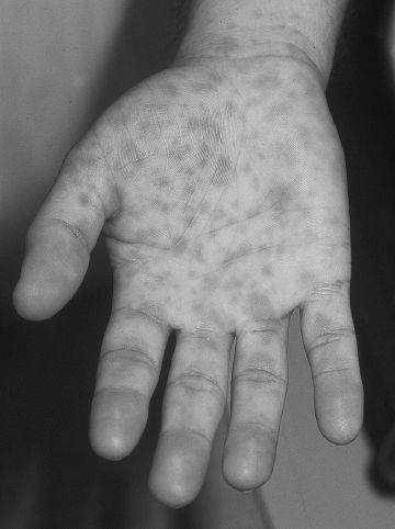
The child’s clinical presentation is most consistent with Rocky Mountain spotted fever. One must have a high index of suspicion because the signs and symptoms during the prodrome are nonspecific. Other findings in children include irritability, severe abdominal pain, conjunctivitis, preseptal edema, and splenomegaly. The rash of Rocky Mountain spotted fever is absent until the third to fifth day of illness. The rash also typically involves the palms and soles and begins on the wrist (see
Figure 105-3
). The rash is the hallmark feature of the disease but may not occur in up to 20% of cases. North Carolina is the most frequent geographic locale for this misnamed disease.

FIGURE 105-3.
Rocky mountain spotted fever. These erythematous lesions will evolve into a petechial rash that will spread centrally. (Reproduced, with permission, from Knoop KJ, Stack LB, Storrow AS, et al. Atlas of Emergency Medicine, 3rd ed. New York: McGraw-Hill; 2010:372. Photo contributor: Daniel Noltkamper, MD.)
10.
(C)
Doxycycline is the drug of choice, even in children younger than 8 years. Tetracycline staining of teeth is dose related and unlikely to occur with a single therapeutic course; doxycycline is less likely than other tetracyclines to stain teeth; and use of the alternative antibiotic chloramphenicol has significant potential toxicity. In addition a retrospective study indicates that chloramphenicol may be less effective than doxycycline for treatment of Rocky Mountain spotted fever.
11.
(B)
In children most cases of ehrlichiosis have been associated with
E chaffeensis
, which causes human monocytic ehrlichiosis. Most human monocytic ehrlichiosis infections occur in people from southeastern and south central United States, but cases of ehrlichiosis have been reported in 48 states. A closely related infection with similar clinical manifestations and course of illness is human granulocytic anaplasmosis, caused by
Anaplasma phagocytophilum.
Pediatric cases have a male predominance, and the peak incidence occurs from May to August. The most common symptoms reported in children include fever, myalgia, and rash. Lymphopenia, thrombocytopenia, and increased serum AST are common.
12.
(E)
Treatment with doxycycline should continue until at least 3 days after defervescence for a minimum of 5-10 days.
13.
(B)
The risk that a neonate will acquire
C trachomatis
is 50% if the mother has a chlamydial infection. The risk for neonatal conjunctivitis is 25-50% and for pneumonia is 5-20%. Pneumonia caused by
C trachomatis
occurs at 2-10 weeks of age. A staccato cough, tachypnea, and rales are characteristic. Wheezing is typically absent. A chest radiograph reveals bilateral interstitial infiltrates and hyperinflation. A diagnostic clue may be the presence of peripheral blood eosinophilia (>400 cells/mm
3
).
14.
(A)
Chlamydia pneumoniae
causes communityacquired pneumonia, prolonged cough illness, and acute bronchitis in children. The organism has been implicated as the cause in 5-10% of community acquired pneumonias in children. The illness tends to have a subacute presentation that is indistinguishable from that caused by
Mycoplasma pneumoniae
. Cough is often prolonged with persistence for 2-6 weeks, and the illness can have a biphasic course. Diagnosis can be confirmed by serologic testing, with the microimmunofluorescent antibody test the most sensitive and specific test available.
15.
(A)
In patients with an underlying immunodeficiency (particularly hypogammaglobulinemia), sickle cell disease, Down syndrome, and chronic pulmonary and cardiac disorders, severe pneumonia with pleural effusion can occur. Infection with
M pneumoniae
has been best described as an influenza-like illness with gradual onset. Many extrapulmonary manifestations have been ascribed to
M pneumoniae
. The detection of
M pneumoniae
DNA by PCR has suggested a role for it in extrapulmonary manifestations such as encephalitis, transverse myelitis, pleural effusion, and bacteremia.
M pneumoniae
has also been implicated as a cause of Stevens-Johnson syndrome.
16.
(A)
The newer macrolide and azalide antibiotics are as effective as erythromycin in achieving clinical and microbiologic cure in children with communityacquired pneumonia caused by
M pneumoniae
. Doxycycline is also effective and can be used for children 8 years and older.
17.
(D)
Rat bite fever in the United States is mainly caused by
S moniliformis
. Nonsuppurative migratory polyarthritis or arthralgias occur in 50% of patients. Generalized adenopathy also occurs. Rat bite fever caused by the spirochete
S minus
manifests with fever and ulceration at the site of the bite. Regional lymphadenitis and lymphadenopathy are associated with the illness.
S minus
infection occurs primarily in Asia.
18.
(E)
Penicillin is the drug of choice for rat bite fever caused by either organism. Alternative drugs include ampicillin, cefuroxime, and cefotaxime. Doxycycline can be used in penicillin-allergic patients who are 8 years of age or older.
 S
S
UGGESTED
R
EADING
Pickering LK, Baker CJ, Kimberlin DW, Long SS.
Red Book
:
2009
Report of the Committee on Infectious Diseases.
28th ed. Elk Grove Village, IL: American Academy of Pediatrics; 2009. Schutze GE, Buckingham SC, Marshall GS, et al. Human monocytic ehrlichiosis in children
Pediatr Infect Dis J.
2007; 26:475-479.