Welcome to Your Child's Brain: How the Mind Grows From Conception to College (5 page)
Read Welcome to Your Child's Brain: How the Mind Grows From Conception to College Online
Authors: Sandra Aamodt,Sam Wang
Tags: #Pediatrics, #Science, #Medical, #General, #Child Development, #Family & Relationships

If the neural tube fails to close fully, miscarriage or birth defects such as spina bifida can result. (In spina bifida, the incomplete closure of the neural tube sometimes leads to the protrusion of the spinal cord from the vertebral column.) Folic acid deficiency in the mother increases the risk of such neural tube defects.
For this reason, women who might become pregnant should take 400 micrograms of folic acid (a B-complex vitamin) every day—or more if they’re having more than one baby. Another source is bread, which in the United States and many other countries is made from folic acid–supplemented flour for this reason. If you are trying to have a baby, you should begin taking this supplement before conception, as many women do not find out they are pregnant until neural tube closure has ended, four weeks after conception.
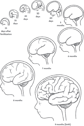
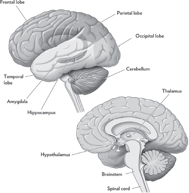
The next stage of development is segmentation, which divides the neural tube into distinct regions by the sixth week of pregnancy. You can think of it as placing walls to define the rooms of a new house, except that segmentation is controlled not by physical barriers but by chemical cues. The largest neural tube region, at the back end of the human embryo, will become the spinal cord. A smaller area at the head end is divided into three regions, which will eventually become different parts of the brain (see figure opposite).
The hindmost of these three regions will become the
brainstem
, which controls mostly subconscious basic functions, such as reflexive movements of the head and eyes, breathing, heart rate, sleep, arousal, and digestion. It also forms the
cerebellum
, which integrates sensory information to help guide movement (for instance, so that you know how forcefully you need to lift your foot when walking).
The middle region will become the brain’s midline structures, including the
hypothalamus
,
amygdala
, and
hippocampus
(see
figure
). The hypothalamus controls many basic processes, such as the regulation of sexual behavior, hunger, thirst, body temperature, and daily sleep/wake rhythms, and the release of stress and sex hormones. Emotions, especially fear, are the responsibility of the amygdala. The hippocampus has two main functions: it stores information into long-term memory, and it is important for spatial navigation.
The third region, at the front of the brain, will become the
thalamus
and cerebral cortex, also called the
neocortex
. Sensory information entering the body through the eyes, ears, or skin travels to the thalamus, in the center of the brain, which filters the information and passes it along to the cortex. Scientists divide the cortex into four parts, or
lobes
. The
occipital lobe
is responsible for visual perception. The
temporal lobe
is involved in hearing, including the understanding of language. This lobe also interacts closely with the amygdala and hippocampus and is important for learning, memory, and emotional responses. The
parietal lobe
receives information from the skin senses, puts together information from all the senses, and directs your attention. The
frontal lobe
generates movement commands, directs the production of speech, and is responsible for selecting appropriate behavior depending on your goals and your environment.
Early in gestation, all these brain regions are tiny. As development continues, chemical markers divide the brain into progressively more regions, defining particular cortical areas, such as those for certain aspects of vision or language. A cluster of cells with a common function is often called a
nucleus
. Once all the brain areas are specified, they grow larger, maturing in sequence from the back of the brain to the front (see
figure
). This process continues through childhood and into adolescence (see
chapter 9
).
The main construction technique in the early stages of brain development is the production of new cells—billions and billions of them. Cells of the early nervous system divide repeatedly to make additional progenitor cells. These cells can even divide as they move, leaving trails of neurons behind them. Cell division
also produces various types of
glial cell
, which contribute to brain function in many ways. One type of glial cell helps guide the placement of neurons early in development by extending long fibers that act as trails for neurons to follow.
The number of cell divisions and what type of cells they produce are tightly regulated by a combination of chemical signals, which vary across brain regions, and interactions with preexisting cells. The addition of new neurons is largely complete by about twenty weeks of
gestational age
(which is counted from the first day of the last menstrual period), or eighteen weeks after conception. A very small number of neurons continue to be generated even into adulthood, and new glial cells are generated throughout life.
During this period, cells are also beginning to differentiate, taking on particular jobs in the brain. Cells differentiate in a series of steps, as their jobs are slowly made more specific by increasingly restrictive chemical signals.
At a basic level, neurons have a lot in common (see
figure
). They receive chemical signals called
neurotransmitters
that are released from other neurons. When neurotransmitter molecules bind to
receptors
on the
dendrites
of the neuron, electrical and chemical signals are generated that can spread—all the way to the cell body in the case of electrical signals. If enough electrical signals occur at the same time, the cell body can make an electrical impulse that is used to talk back to other neurons.
This output signal, called an
action potential
or spike, is conveyed down the
axon
, a very long, thin fiber that reaches from the brain to the target, as far away as the toe in some cases. Each neuron has a single axon, which often branches to reach multiple targets. Neurotransmitter molecules are contained in specialized areas at the ends of the axonal branches and released by the arrival of a spike. When a neurotransmitter binds to receptors on another neuron’s dendrites, that target neuron may be electrically excited or inhibited, depending on the identity of the neurotransmitter. The point of connection between axon and dendrite is called a
synapse
. Final stages of differentiation often depend on neurons’ interactions at synapses.
Glia also come in different flavors. Some glia wrap themselves around axons like the insulating plastic sheath on electrical wire, forming a layer called
myelin
to increase the speed of neural communication. Other glia line blood vessels to control which chemical signals are permitted to pass into and out of the brain. Still others form the brain’s defense system, engulfing and removing foreign
matter and debris from dying cells. Glia too become differentiated by exposure to chemical signals, generally a bit later than the neurons in the same areas.
The first step in the wiring process occurs before birth, as these billions of neurons extend axons toward their targets. Fortunately, distances are much shorter in the fetus than they would be in an adult. It also helps that brain tissue is less crowded than it will eventually become, just as it’s easier to run electrical wires and plumbing in a house before the interior walls have been put up. Only the earliest-arriving axons must find their way by themselves, navigating via chemical signals or by finding particular guidepost cells.
Later axons extend along the pathways laid down by these early pioneer axons, just as you might guide a new wire through a bundle of previously installed wires, except that the new axon is actually being created as it progresses. A bundle of axons in the brain is called a
nerve
. A region at the tip of the elongating axon called the
growth cone
samples the environment within the brain in different directions by extending and retracting small protrusions, making it look as though the growth cone is sniffing out the correct path. Depending on their identity, these chemicals may either attract or repel the growth cone. Some can even cause it to abruptly change its responsiveness to other molecular cues, a form of sophisticated navigational logic.
Once an axon has found its approximate destination in the brain, it must pinpoint its target cells from among millions of candidates. This process starts with molecular cues that tell the axon to slow down and start exploring an area whose boundaries may be marked with a repellent signal to prevent the axon from exiting. Some brain areas help the axon to navigate by providing a local map, in which the concentration of a chemical signal (or several) descends steadily across the area. Other areas use a large number of related
proteins
to mark local position so axons can find their way to the right neurons. Proteins are the universal building blocks made by cells for a wide variety of functions. In this case, the function is to say to an axon, “You are here.”
PRACTICAL TIP: LESS STRESS, FEWER PROBLEMS

Next time you’re stressing about your future child, ask yourself whether this stress is really necessary. Neuroscientists are able to discover what stress does by studying its effects on laboratory animals. Maternal stress increases the risk of a variety of problems, including cleft palate, depression-like behavior, a touchy stress-response system in adulthood (see
chapter 26
), and attention deficits and distractibility (see
chapter 28
). Stress hormones released by the mother animal act on the fetus directly and also reduce the placenta’s ability to protect the fetus from these hormones in the future.
Because it would be highly unethical to stress pregnant women deliberately, most research in people has relied on looking for correlations, which is less reliable than experimental results (see
Did you know? Epidemiology is hard to interpret
). Some recent studies have examined children born after their mothers experienced natural disasters during pregnancy. This type of study comes as close as is ethically possible to randomly placing women into stressed and unstressed groups.
One group of researchers identified all tropical storms or hurricanes that hit Louisiana between 1980 and 1995 and then determined how many autistic children in the records of the state health system had been in the womb when their mother’s home was hit by one of these storms. The risk of autism was significantly higher for children whose mothers had been stressed during pregnancy—though most cases of autism probably result from other causes (see
chapter 27
).
By scientific standards, this evidence is far from ironclad, but there are two reasons to believe it’s not mere chance. First, the incidence of autism was higher only for those children whose mothers were in the fifth, sixth, or ninth month of pregnancy at the time of the hurricane, suggesting that there is a period when the effects of stress on development are long-lasting (see
chapter 5
). Second, children whose mothers were exposed to more severe storms had a higher risk of autism than children whose mothers were exposed to less severe storms. This research will need to be replicated before we can consider it definitive, but it does suggest that prenatal stress may increase the chances of autism.